Japanese
English
- 有料閲覧
- Abstract 文献概要
- 1ページ目 Look Inside
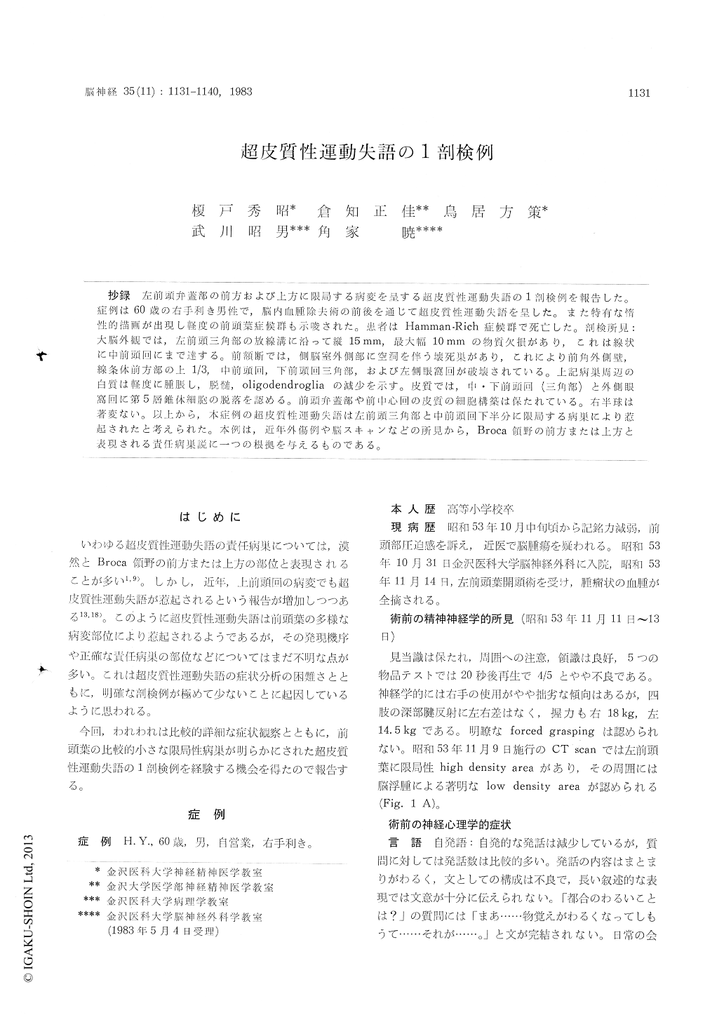
抄録 左前頭弁蓋部の前方および上方に限局する病変を呈する超皮質性運動失語の1剖検例を報告した。症例は60歳の右手利き男性で,脳内血腫除去術の前後を通じて超皮質性運動失語を呈した。また特有な惰性的描画が出現し軽度の前頭葉症候群も示唆された。患者はHamman-Rich症候群で死亡した。剖検所見:大脳外観では,左前頭三角部の放線溝に沿って縦15mm,最大幅10mmの物質欠損があり,これは線状に中前頭回にまで達する。前額断では,側脳室外側部に空洞を伴う壊死巣があり,これにより前角外側壁,線条体前方部の上1/3,中前頭回,下前頭回三角部,および左側眼窩回が破壊されている。上記病巣周辺の白質は軽度に腫脹し,脱髄,oligodendrogliaの減少を示す。皮質では,中・下前頭回(三角部)と外側眼窩回に第5層錐体細胞の脱落を認める。前頭弁蓋部や前中心回の皮質の細胞構築は保たれている。右半球は著変ない。以上から,本症例の超皮質性運動失語は左前頭三角部と中前頭回下半分に限局する病巣により惹起されたと考えられた。本例は,近年外傷例や脳スキャンなどの所見から,Broca領野の前方または上方と表現される責任病巣説に一つの根拠を与えるものである。
An autopsy case of transcortical motor aphasia is presented with a pathology located anterior and superior to the pars opercularis of the left inferior frontal gyrus.
Case H. Y. A 60-year-old right-handed man.
On Nov. 14, 1978, the patient had surgery to remove cerebral hematoma in the left frontal lobe. In the neuropsychological examination before the operation, he had shown the clinical features of transcortical motor aphasia characterized by good comprehension of language, preserved repetition, and spontaneous speech disorder.In this stage, it was supposed that the underlying disturbance of spontaneous speech was due to the disabilities of contextual constructions of sentences rather than the lack of speech initiation. Following the opera-tion, however, spontaneous speech disappeared completely for several days. At the same time, the patient showed problems in comprehension, reading, writing and confrontation naming as well as symptoms of disorientation, pathological inertia and 'loss of initiation' in the psychomotor domain. During the following three months, however, the patient did show slight improvement, except for contextual sentence constructions and pathological inertia when taking the complex animal drawing test. In his terminal stages, the clinical symptoms could be summarized as transcortical motor aphasia and mild frontal lobe syndrome. On march 1, 1979, the patient died of Hamman-Rich syndrome.
Postmortem examination : The brain weighed 1294 gm. The external observation of the brain disclosed the linear tissue defect, abou 15 mm in length and 10 mm in width, along the radial sulucus of the pars triangularis of the left inferior frontal gyrus. Coronal sections revealed a necrotic lesion involving the gray and white matter of the pars triangularis of the inferior frontal gyrus and the white matter underlying the lower half of the middle frontal gyrus in the left hemisphere. Mic-roscopic examination : The serial sections of ceroidin-embedded materials were stained withthe method of Woelcke, Kluver-Barrera, hae-matoxylin and eosin, Nissl and Holzer. In the left hemisphere, there was a cavitated necrosis in the following regions ; the middle frontal gyrus, the pars triangularis of the inferior frontal gyrus, the lateral side of the orbital gyrus, the lateral wall of the anterior horn of the lateral ventricle, and the top third of the anterior striatum. The white matter surrounding the lesion was slightly swol-len, showing demyelination and a decreased popu-lation of oligodendroglia but no fibrillary gliosis. There was a decreased number of pyramidal cells in the 5th layer of some parts of the middle frontal gyrus, the pars triangularis of the inferior frontal gyrus, and the lateral side of the orbital gyrus. The cytoarchitecture and number of neurons werewell preserved in the pars opercularis of the inferior frontal gyrus and the precentral gyrus. There were no pathological findings in the right hemisphere.
Based on these findings, it was concluded that the clinical features of transcortical motor aphasia in this patient were due to the localized lesion in the pars triangularis of the left inferior frontal gyrus and the lower half of the middle frontal gyrus. This conclusion supports the recent auge-ment, based on observations of head trauma cases and the findings of brain scans, that many cases with transcortical motor aphasia have the patho-logy located either anterior to or superior to Broca's area.

Copyright © 1983, Igaku-Shoin Ltd. All rights reserved.


