Japanese
English
- 有料閲覧
- Abstract 文献概要
- 1ページ目 Look Inside
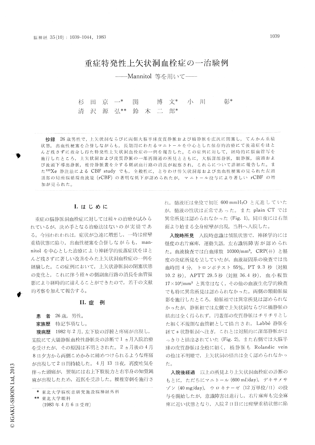
抄録 26歳男性で,上矢状洞ならびに両側大脳半球皮質静脈および橋静脈を広汎に閉塞し,てんかん重積状態,出血性梗塞を合併しながらも,長期間にわたるマニトールを中心とした保存的治療にて後遺症をほとんど残さずに救命し得た特発性上矢状洞血栓症の一例を報告した。この症例に対して,経時的に脳血管写をを施行したところ,上矢状洞および皮質静脈の一部再開通の所見とともに,大脳深部静脈,眼静脈,前頭および後頭下導出静脈,椎骨静脈叢を介する側副血行路の消長が観察され,これらについて詳細に報告した。また133Xe静注法によるCBF studyでも,全般性に,とりわけ傍矢状洞部および出血性梗塞の見られた左頭頂部の局所脳循環血流量(rCBF)の著明な低下が認められたが,マニトール投与により著しいrCBFの増加が見られた。
A cured case of superior sagittal sinus throm-bosis is reported. The patient, a 26-year-old man, also displayed severe complications such as hemor-rhagic infarction and status epiiepticus. Although his prognosis was considered to be extremely poor, conservative treatment, with mannitol, ste-roid, anticonvulsants, etc. was effective, and hewas discharged without any neurological deficit.
This report discusses the clinical course, CT findings, angiographical findings and regional ce-rebral blood flow (rCBF).
At first, CT showed no abnormal findings, but hemorrhagic infarction was detected on the 3rd day after the onset. Follow-up CT showed subcor-tical low density area, hemorrhagic infarction with perifocal brain edema, midline shift etc ; the focus of hemorrhagic infarction was almost absorb-ed 2.5 months after the onset.
Cerebral angiogram showed not only the obs-truction of the superior sagittal sinus but also that of cortical veins of cerebral convexity at first. Follow-up angiogram showed the development of collateral circulations such as deep cerebral, op-thalmic and emissary veins.
On CBF study, low rCBF at the bilateral para-sagittal region was observed, but marked increase of rCBF was measured in the parasagittal region, especially at the site of hemorrhagic infarction after the administration of 20% mannitol. We, therefore, consider mannitol as an effective agent for the treatment of cerebral sino-venous throm-bosis.

Copyright © 1983, Igaku-Shoin Ltd. All rights reserved.


