Japanese
English
- 有料閲覧
- Abstract 文献概要
- 1ページ目 Look Inside
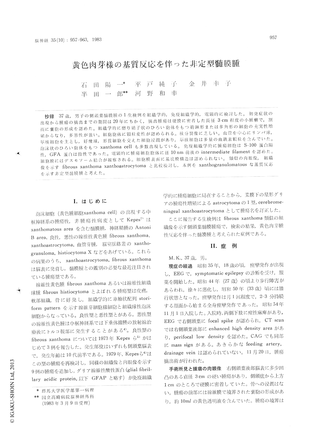
抄録 37歳,男子の側頭葉髄膜腫の1生検例を紐織学的,免疫組織学的,電顕的に検討した。初発症状の出現から腫瘍の摘出までの期間は20年にちかく,摘出腫瘍は硬膜に密着した長径3cm程度の小腫瘤で,割面に嚢胞の形成を認めた。組織学的に磨り硝子状のひろい胞体をもつ紡錘形または多角形の細胞の充実性増殖からなり,多態性が強い。細胞胞体に顆粒変性が認められる。核分裂像に乏しい。血管を中心にリンパ球,単核細胞を主とし,好酸球,形質細胞を交えた細胞浸潤があり,単核細胞は多量の血鉄素顆粒を含んでいた。泡沫状のひろい胞体をもつxanthoma cell 多数出現している。免疫組織学的に腫瘍細胞はS−100蛋白陽性,GFA蛋白は陰性であった。電顕的に腫瘍細胞胞体に径10nm前後の intermediate filamentを認めた。細胞膜にはデスモソーム結合が観察される。細胞膜表面に基底膜構造は認められない。類似の肉眼像,組織像を示すfibrous xnthoma xanthoastrocytomaと比較検討し,本例をxanthogranulomatousな基質反応を示す非定型髄膜腫と考えた。
A case of atypical meningioma with xanthogra-nulomatous stromal reaction was described in a 37 year-old man. At the age of 18, the patient started to have seizures. He became aware of disturbances in gait since 27 years of age. At the age of 37 he came to the hospital for detailed examinations and adequate treatment. EEG show-ed focal spikes at right temporal area and the CT scan disclosed abnormalities indicating the presence of a right posterior temporal mass.
At craniotomy, a tumor was found located over the surface of right temporal lobe. It was well-circumscribed, firm in consistency and was attatch-ed to the dura, measuring 3cm in its greatest diameter. Gross total removal was accomplished. On cut section, the tumor was greyish in color mottled variously with yellow or rustbrown tint. It contained a cyst.
Microscopically, the tumor was made up of compact masses of spindle-shaped or polygonal cells. The cells were arranged in areas in inter-lacing fascicles or forming whorles. In many places the tumor showed pleomorphic cytology. Mitotic figures were found few in number. The cytoplasm of the neoplastic cells appeared groundglass-like. Some of them contained hyaline globules. The globules were found to react positive with P. A. S. technique, and this procedure was resistant against diastase digestion. They were also stained positive with P. T. A. II. stain. In some areas foamy xan-thomatous cells were found in groups. A large number of lymphocytes, plasma cells, eosinophiles and macrophages ingesting brownish pigments were found infiltrating around vascular vessels. Berlin blue stain disclosed the pigments to be iron-positive. Some tumor cells contained also iron-positive pigments. Reticulin fibrils were found almost confined to the perivascular areas where numbers of inflammatory cell infiltrates were seen. PAP immunoperoxidase method demonstrated the presence of S-100 protein in the cytoplasm of neoplastic cells. The GFA protein was not demon-strable in any of the cells. Xanthoma cells were found S-100 protein-negative, showing a sharp contrast to positively stained neoplastic cells.
Electron microscopic studies were made on the formalin-fixed tumor tissue. The neoplastic cells were characterized by the presence of intermediate filaments 8-10 nm in width in the cytoplasm and well-developed desmosomal junctional devices between two opposed cell membranes. Two types of cytoplasmic inclusions were found in occasional neoplastic cells. The one was electron dense par-ticles of irregular shape which were composed oflarge collections of presumed ferritin particles, 80-100A in size, and the other was membrane-bound osmiophilic granules of diverse internal structure, 0.2-1.7 μ in size. The former structure was thought to be siderosome and the latter to be equated with P. A. S. positive hyaline granules in light microscopy. Foamy cells were found in electron microscopy as cells loaded with lipid vacuoles of varying size.
Clinical manifestations with indices of slow tumor growth, location, gross and microscopic characteristics of this neoplasm resembled thoseof cases reported by Kepes et al. as xanthoastrocy-toma. However, the presence of intracytoplasmic intermediate filaments, well-developed desmosomal junctional devices and the absence of basement membrane over the surface of individual tumor cells in electron microscopy, and a negative immunohistochemical demonstration of GFAP seemed to favor the diagnosis of meningioma.
From these observations this case was summar-ized as a meningioma of unusual histology with xanthogranulomatous stromal reaction.

Copyright © 1983, Igaku-Shoin Ltd. All rights reserved.


