Japanese
English
- 有料閲覧
- Abstract 文献概要
- 1ページ目 Look Inside
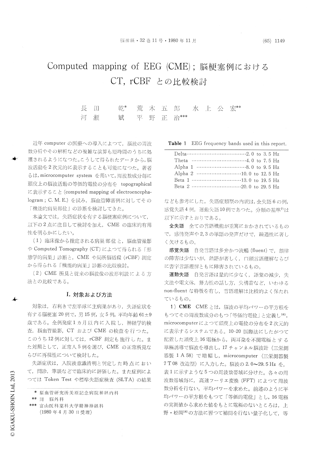
近年computerの医療への導入によつて,脳波の周波数分析やその解析などの複雑な演算も短時間のうちに処理されるようになつた。こうして得られたデータから,脳波活動を2次元的に表示することも可能になつた。著者らは,microcomputer systemを用いて,周波数成分毎に頭皮上の脳波活動の等価的電位の分布をtopographicalに表示すること(computed mapping of electroencepha—logram;C.M.E.)を試み,脳血管障害例に対してその「機能的病巣部位」の診断を検討してきた。
本論文では,失語症状を有する脳梗塞症例について,以下の2点に注目して検討を加え,CMEの臨床的有用性を明らかにしたい。
Computed mapping of electroencephalogram (CME) is a newly developed microcomputer system to display equipotential maps of square roots of powerspectra over each frequency band on color cathode ray tube (CRT). This system was employed in examinations of twenty aphasics due to cerebral infarction incomparison with CT and regional cerebral blood flow (rCBF) study. In this system, data from the scalp were amplyfied via 17-channel EEG polygraph (San-ei 1A58) and connected with on-line microcomputer (San-ei 7T08). Spectral analysis was carried out using fast Fourier trans-formation (FFT) technique for each channel. The square roots of powerspectra were defined as equi-valent potentials over each frequency band. Ac-cording to Ueno and Matsuoka's method interpola-tion was performed and interpolated values were quantitized into eleven steps which were represented in eleven colors respectively on color CRT in each frequency band ; delta, theta, alpha-1 (slow alpha),alpha-2 (fast alpha), beta-1 and beta-2. Regional CBF study was performed as to the left hemis-pheres on twelve patients by 133Xe intracarotid injection technique using 16-channel scintillation detector and on-line microcomputer (Meditronic Cerebrograph). In a control group of five normal volunteers, alpha activity was high and symmetrical and there was no particular high voltage focus in slow component except for the frontal delta foci representing blinks which were confirmed by EEG inspection. As regards reproducibility of the re-sults, an analysis of 120 second EEG data appeared to be satisfactory in comparison with analyses of various periods of EEG data. Six patients had global aphasia, four had sensory and ten had motorrespectively. In five of global aphasics high volt-age areas were found predominantly in the left side in delta band which corresponded to the lesions on CT, and in another global aphasic, delta activity was high bilaterally. Regional CBF study revealed left hemispheric severe ischemia in three cases of global aphasics. In three of sensory aphasics alpha activity was confined to the right temporo-occipital region and there was no high voltage focus in slow component. Another cases of sensory aphasia showed a high voltage focus round the left temporal region which well corresponded to the lesion on CT. Regional CBF study demonstrated a marked ischemia of the left temporal lobe. In this case a functional lesion provided by CME and rCBF well corresponded to the morphological lesion on CT. Five motor aphasics showed asymmetry of alpha distribution lateralized to the right side and no obvious locali-zation of slow component and three showed delta activity lateralized to the left side and alpha activity to the right side. In one case of motor aphasia, there was no obvious low density area which likely caused motor aphasia on CT, but slow component was prominent in the left side and alpha activity was relatively lateralized to the right side on CME. In this case rCBF study revealed left hemispheric severe ischemia. Accordingly it can be said thatCME and rCBF study revealed left hemispheric dysfunction which was not suggested by CT. In conclusion, CME described in this report has three advantages, the first is to detect the localization of high voltage area of slow component topographically and semi-quantitatively. The second is to demonst-rate the distribution of alpha activity topographi-cally and objectively. The third is to provide functional lesions even in absence of morpho-logical lesion on CT. As regards functional lesions of the brain, using a microcomputer system such as CME, we can obtain topographical, semi-quanti-tative and objective aspects of electrophysiological activity of the brain though the source of the data is the same as conventional EEG interpretation.

Copyright © 1980, Igaku-Shoin Ltd. All rights reserved.


