Japanese
English
- 有料閲覧
- Abstract 文献概要
- 1ページ目 Look Inside
1.はじめに
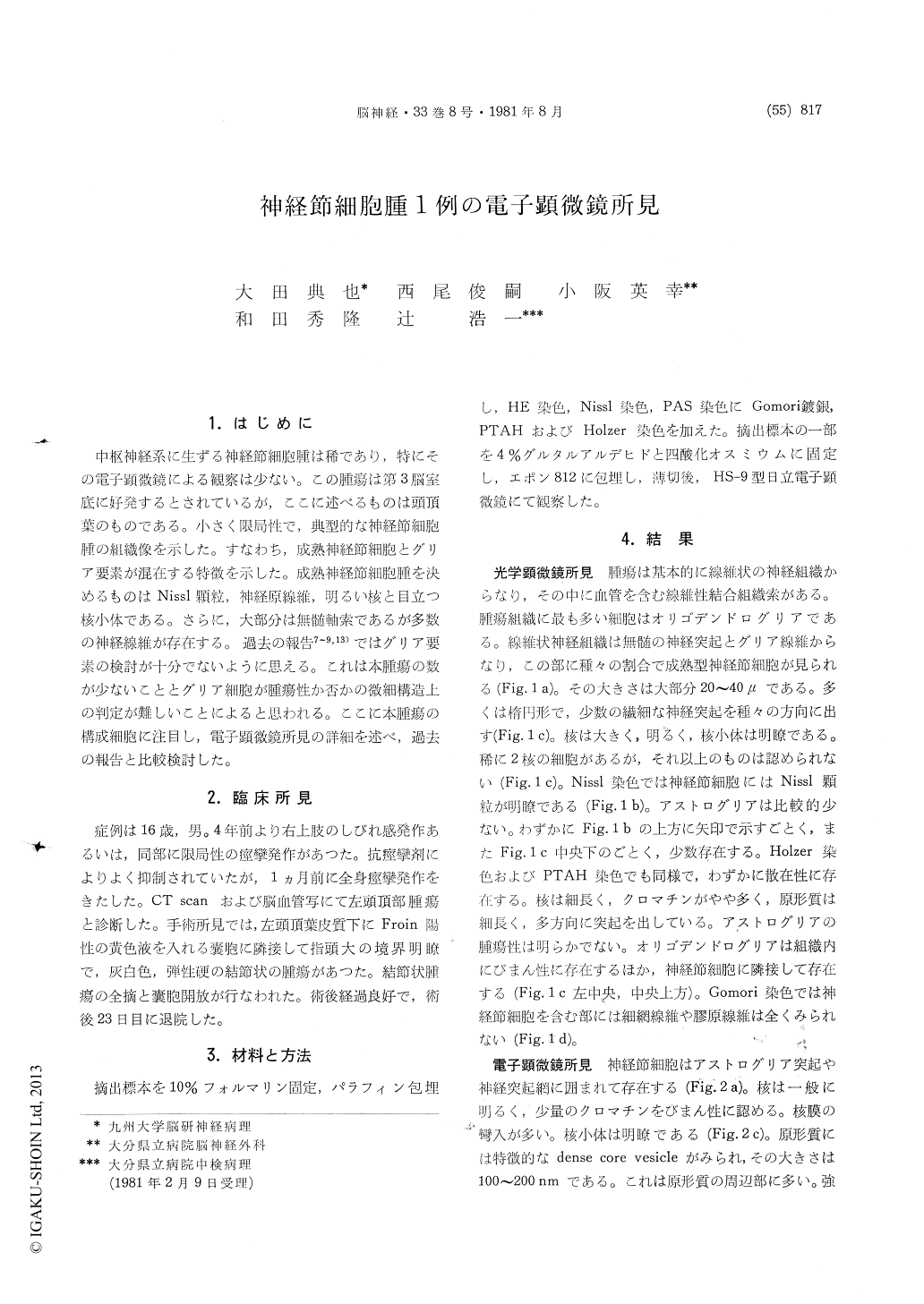
中枢神経系に生ずる神経節細胞腫は稀であり,特にその電子顕微鏡による観察は少ない。この腫瘍は第3脳室底に好発するとされているが,ここに述べるものは頭頂葉のものである。小さく限局性で,典型的な神経節細胞腫の組織像を示した。すなわち,成熟神経節細胞とグリア要素が混在する特徴を示した。成熟神経節細胞腫を決めるものはNissl顆粒,神経原線維,明るい核と目立つ核小体である。さらに,大部分は無髄軸索であるが多数の神経線維が存在する。過去の報告7〜9,13)ではグリア要素の検討が十分でないように思える。これは本腫瘍の数が少ないこととグリア細胞が腫瘍性か否かの微細構造上の判定が難しいことによると思われる。ここに本腫瘍の構成細胞に注目し,電子顕微鏡所見の詳細を述べ,過去の報告と比較検討した。
The tumor tissue removed surgically from the left parietal subcortical area of a 16-year-old boy was studied by light and electron microscopy.
Our results were identical to cases reported previ-ously. By light microscopy, the neoplasm was composed mostly of mature and immature ganglion cells and glial cells. Ganglion cells with large nuclei and prominent nucleoli had characteristic Nissl substance in various amounts and they were encompassed in a network of fibrous connective tissue. Glial cells were mainly oligodendrocytic. Astrocytes were small in number and did not show neoplastic growth. By electron microscopy, mature ganglion cells contained dense core vesicles in their cytoplasm and processes. The size of the vesicles ranged from 100 to 200nm. Furthermore ganglion cells were surrounded by their own processes and also processes of glial cells. Ganglion cells were rarely found in synaptic contact with adjacent processes. Instead, oligodendrocytes were sometimes situated adjacent to these ganglion cells. These findings suggest that the tumor under study may be of hamartomatous origin. Aside from mature ganglion cells and glial cells, immature cells were present. From viewing the architecture of these immature cells, we concluded that they were also ganglion cells.

Copyright © 1981, Igaku-Shoin Ltd. All rights reserved.


