Japanese
English
- 有料閲覧
- Abstract 文献概要
- 1ページ目 Look Inside
I.はじめに
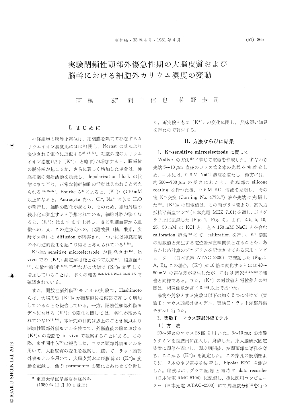
神経細胞の膜静止電位は,細胞膜を隔てて存在するカリウムイオン濃度比にほぼ相関し,Nernstの式により決定される電位に近似する35,36,37)。細胞外腔のカリウムイオン濃度(以下〔K+〕0と略す)が増加すると,膜電位の脱分極が起こるが,さらに著しく増加した場合は,神経細胞の発射活動を誘発し,depolarization blockの状態にまで至り,正常な神経細胞の活動は失われると考えられる35,36,37)。Bourkeら3)によると,〔K+〕0が10mM以上になると,Astrocyte内へ,Cl—,Na+さらにH2Oが移行し,細胞の膨化が起こり,そのため,細胞外腔の狭小化が発生すると予想されている。細胞外腔が狭くなると,〔K+〕0はますます上昇し,さに毛細血管から組織への,又,この逆方向への,代謝物質(糖,酸素,炭酸ガス等)のdiffusionが阻害され,ついには神経細胞の不可逆的変化も起こり得ると考えられている3,18)。
K+—ion sensitive microelectrodeが開発され45),invivoでの〔K+〕0測定が可能となつて以来23),脳虚血16,19),拡散性抑制5,6,36,37,41)などの状態で〔K+〕0が著しく増加していることは,多くの報告2,3,5,6,9,16,17,23,36,37)で確認されている。
The high concentration of extracellular potas-sium ((K+)O) impedes neuronal activity by depolariz-ing the membrane potential and further causing depolarization block or conduction block, and also causes swelling of astrocytes, which may result in narrowing of extracellular space and affect the diffusion of metabolites. Thus, when high con-centration of extracellular potassium prolongs, pa-thological changes of neurons may ensue. In such state as cerebral ischemia, contusion or spreading depression, marked elevation of extracellular potas-sium was reported by many authors. In order to reveal the changes of extracellular potassium dur-ing the acute phase of closed head injury, follow-ing experimental studies were performed.
In the first experiment, closed head injury model of the mouse was used. Directly after the impact with 600g.cm, spontaneous movement of the animal disappeared for about 5-6 minutes often accompa-nied by immediate epilepsy. In this state, cortical (K+)0 increased from the control level of 4.1±1.8mM to 20-30mM. The elevated (K+)0recovered gradually within 10-30 minutes. Directly after the same impact, apnea, bradycardia and low voltage EEG also appeared.
In the second experiment, closed head injury model of the rat was used. With the impact of 9000 g. cm, the same concussion-like symptoms ap-peared as those of mice. Directly after the impact, (K+)0 in the cortex and brain stem elevated but the change patterns of cortical (K+)0 and brain stem (K+)0 were different. The different patterns in changes of (K+)0 were classified into four types.
Type 1. Both cortical and brain stem (K+)0ele-vated significantly immediately after the trauma, and recovered to control level within 30 minutes. Type 2. Both (K+)0 elevated significantly as in type1 but only brain stem (K+)0 recovered within 30 minutes. Type 3. With the exacerbation of vital signs, both (K+)0 elevated over 50mM, resulting in the death of the animal. Type 4. Although corti-cal (K+)0elevated significantly after the impact, the increase of brain stem (K+)0 was below 10mM. In type 1,2 and 3, blood pressure always elevated and brady- or apnea appeared immediately after the impact. In type 4, blood pressure after the impact showed to become slightly lower transiently as com-pared to control blood pressure.
In control study, spreading depression was elic-ited by application of KCl-solution on the cor-tex. Spreading depression caused the elevation of cortical (K+)0 only, and almost no changes of brain stem (K+)0 were observed. The present study sug-gested that during the acute phase of closed head injury, diffuse neuronal disturbances involving the brain stem existed. And the elevation of brain stem (K+)0 is an important finding of this study because it may affect the level of consciousness directly by the mechanisms as described above.

Copyright © 1981, Igaku-Shoin Ltd. All rights reserved.


