Japanese
English
- 有料閲覧
- Abstract 文献概要
- 1ページ目 Look Inside
I.はじめに
Foramina parietalia permagnaは,頭頂骨の後上部において,矢状縫合をはさんでほぼ左右対称に存在する頭頂骨の局所的骨欠損である。
1707年,Lancisiの報告が最初であると言われ,1922年GoldsmithのCatlinなる家系の報告以来本症が遺伝疾患と考えられるようになつたが,現在までに世界で約200例の報告にとどまるまれな疾患である。
Foramina parietalia permagna are usually paired, bilaterally symmetrical bone defect at the para-sagittal area of the posterior parietal bone. In 1707 Lancisi was the first to report this anomaly in postmortem examination. In 1922 Goldsmith presented hereditary incidence of the malformation in the Catlin family. He found this anomaly in 16 members (13 males and 3 females) in five generations of the family and called it the 'Catlin mark '. Since then, several families in which the anomaly occurred have been published. Formal mode of genetic transmission remains unknown. This condition has been considered rare and about 200 cases have been reported from many parts of the world. We present this malformation seen in five members in three generations of a family and review the literature.
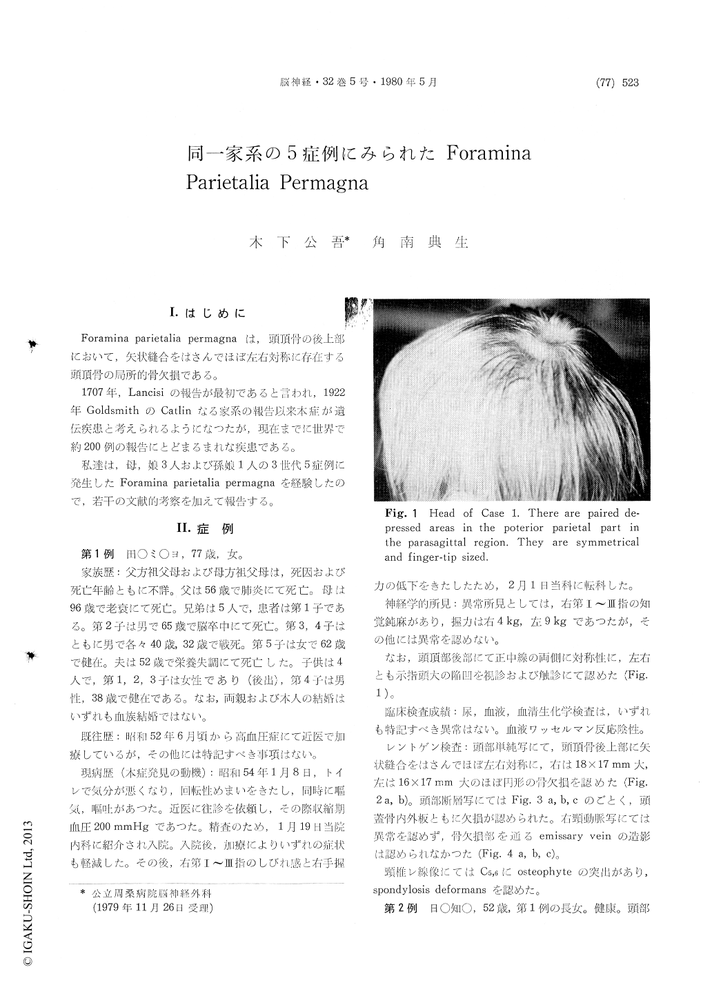
Case 1, a 77-year-old woman, was admitted on January 17, 1979 with a complaint of vertigo, nausea and vomiting. These symptoms gradually improved but she developed some numbness in the right first, second and third fingers. Physical ex-amination revealed two depressed areas in the scalp, one on each side of the midline in the posterior parietal region. Skull x-ray films showed two round bone defects, the right one being 18×17 mm in diameter and the left one 16×17 mm. The laminograms revealed that the bone defect involved the outer and inner tables. A right carotid angiography demonstrated no abnormal findings. No emissary vein was visualized. Family history disclosed that the similar bone defects were found in Case 2 (the first daughter of Case 1, aged 52), Case 3 (the second daughter of Case 1, aged 47), Case 4 (the third daughter of Case 1, aged 42) and Case 5 (the first daughter of Case 4, aged 17).
In 1919 Pamperl entitled this anomaly foramina parietalia permagna. This anomaly seems to be extremely rare in Japan. Okuda et al. (1965) first reported a familial incidence of this disorder affecting mother and her daughter. Our presen-tation is the second one with term of familial occurrence.
Foramina parietalia permagna are usually paired and are located symmetrically in the parietal bone on each side of the posterior one-third of the sagittal suture. In this area there are normal bone defect called foramen parietale, which contains emissary veins. The diameter of the parietal foramina ranges from 0.5 to 2.0 mm.
The parietal bone ossifies from two center in membrane in the parietal eminence at about the seventh or eighth week of fetal life. Ossification proceeds in a radial fashion toward the sagittal suture and ceases in the area adjacent to the parietal foramina. Extensive defective ossification in the medial aspect of the parietal bone is con-sidered to result in foramina parietalia permagna. In infancy this anomary begins as a single large midline defect of the parietal bones. Subsequently the large defect is divided into two parts by ex-tensions of the parietal ossification into the medial aspect of the parietal bones.
From a review of published cases, males and females are equally affected. It is of interest that all of the 7 patients reported in Japan including our cases are all females. The foramina parietalia permagna is usually not neurologically significant but must be distinguished from a number of dis-orders producing bone destruction. Cranioplasty has been recommended if these foramina are more than 3 cm in diameter in children beyond three years of age.
Copyright © 1980, Igaku-Shoin Ltd. All rights reserved.