Japanese
English
- 有料閲覧
- Abstract 文献概要
- 1ページ目 Look Inside
I.はじめに
近年脳あるいは心臓疾患の手術に際し,各種抗生剤や副腎皮質ホルモンが,大量に投与されるようになつてから,いわゆる菌交代現象や,免疫反応の低下に伴う12),真菌性脳病変の増加傾向が注目されるようになつた13)。
脳にみられる原発性または転移性の真菌性肉芽腫は極めて稀であり,とりわけ脳のchromomycosisは外国では1948年Bonneら2)の報告を嚆矢に,また本邦では福代4),笹野ら16)の報告に接して以来,著者の調べ得た限りでは,現在まで28例をみるに過ぎない。
This is the autopsy report of a case of chromo-mycosis developing on the skin, left lung, brain, lympho nodes, liver, ileocecal region and kidney of a 15 year-old Japanese boy.
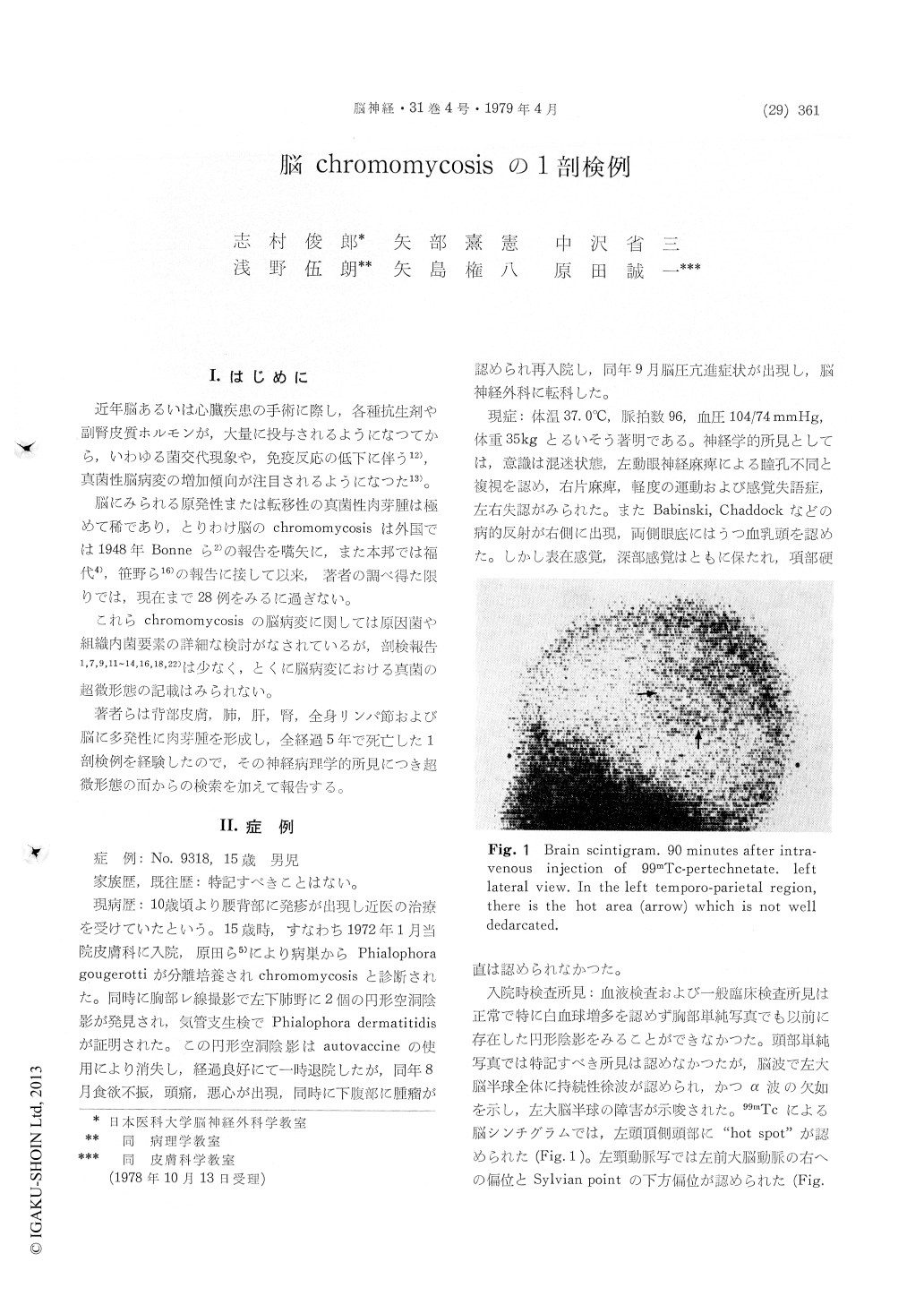
Clinical findings. : A 15 year-old male, with a five-year history of complaints of skin eruptions on his back was hospitalized in January, 1972. Skin biopsy was performed and a positive culture for phialophora gougerotti was obtained. He com-plained of nausea, vomiting and headache in August, 1972 and was reffered to department of neuro-surgery in our hospital. Neurological examination revealed motor and sensory aphasia, chocked disc and right hemiparesis. The cerebral scintigram showed large spot in the left temporo parietal area. The cerebral angiogram revealed space occupying lesions in the left cerebral hemisphere. On September 21, 1971 craniotomy was performed and a brown partially reddish colored granuloma of Ca 40 g. weight was extripated. Although the patient showed some improvement with the ad-ministration of autogenous vaccine, he died on November 27, 1972.
Autopsy findings. : Brain weight 2,000 gr. A postmorten examination revealed irregular shapedand multiple mycotic granuloma with yellowish brown color at the lentiforme nucleus, internal capsule, claustrum, corpus callosum, septum pellucidum, cerebellum and pons on the left side, and bilateral white matter of parietal lobe. In addition, black nodule with a few millimeter in diameter were disseminated on the cerebral basis especially cerebello medullary cistern.
Culture findings phialophora gougerotti was isolated from the skin lesions on the back and left upper arm, and phialophora dermatitidis was also isolated from the left lung, brain, lymph nodes and kidney.
Microscopic findings of the brain. : Microscopy show massive infiltration of small round cells and coagulation necrosis in the granuloma and there are many septate hyphae in the coagulated necrotic tissues and multinucleated giant cells surrounding the blood vessels. Almost all mycotic fungi of the cerebral granuloma are of hyphae type and not spore type.
In ultrastructual findings, the mycotic fungi have fibrous fibril around the cell wall. Hyphae contains many vacuoles, mitochondria, and septum of cyto-plasm. For the purpose of recognizing polysac-charide compound, section were stained with the periodic acid methenanine Silver Stain according to Yajima's method in 1968. And PAM positive granules are visible by this method in the cell membrane of the mycotic fungi. From these findings it can be said that the cerebral fungi as well as skin fungi have polysaccharide compound in the membrane.
We found 10 autopsy reports of the cerebral chromomycosis in literature. These autopsy cases showed that the most marked pathological changes were located in the cerebral white matters and those the next remarkable changes was seen in the extramedullary part of the cerebrum.
Copyright © 1979, Igaku-Shoin Ltd. All rights reserved.