Japanese
English
- 有料閲覧
- Abstract 文献概要
- 1ページ目 Look Inside
I.はじめに
いわゆる嚢状脳動脈瘤の成因,発育,破裂機序については先人により流体力学的な立場,あるいは病理組織学的な面からの検討が種々行われており,なかでも成因に関してはForbus9)に代表される先天説,Crawford5),Stehbens18)による後天説およびCarmichael4)等の先天,後天両者折衷説等と議論が多い。一方発育,破裂に関しての検討は少く僅かにFerguson8),Jain13)等により流体力学的面からの考察が行われている程度である。
しかし,いずれの場合にも多くは破裂前の脳動脈瘤についてその成因,成長および破裂の検討を行つたものであり,一旦破裂した後の脳動脈瘤の修復およびその後の経時的変化を検討した研究は極めて少ない7,15,18)。さて実際臨床面では脳動脈瘤が破裂を契機として明らかに成長したと考えられる症例に遭遇することがあり,従来の成長機序のみでは説明できないこともしばしば経験される。この点から著者らは嚢状脳動脈瘤の発生,破裂,修復および発育機序を更に解明する目的で臨床経過を軸とした脳動脈瘤の病理組織学的検索を行つたので報告し,特に脳動脈瘤の破裂後の修復およびその後の発育機序について考察を加える。
The origin and mechanism of rupture, repair andgrowth of intracranial saccular aneurysms werestudied in this paper.
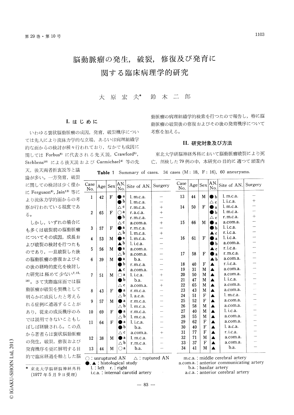
Forty-five aneurysms, 23 unruptured and 22ruptured aneurysms, of 60 aneurysms found from34 brain specimens with aneurysm rupture, wereinvestigated from the clinical and histopathologicalviewpoints.
Nineteen of 23 unruptured aneurysms weresmaller than 4mm in diameter. In these unrupturedaneurysms, walls of aneurysms smaller than 3mmin diameter were mainly composed of endothelialcells and fibrous tissue. Walls of aneurysms largerthan 3mm were irregular in thickness and col-lagenous in some portions. Walls of aneurysmslarger than 4 mm were mainly collagenous andextremely thin portions were observed in theirdomes.
Twenty-two ruptured aneurysms were all largerthan 4mm in diameter and rupture points wereall in the domes. Sixteen cases died within 3weeks after the initial hemorrhage. Histologically,at the rupture point, a bulging protective layerwas formed extending from the original aneurysmalwall. Reruptures occurred in the weak point ofthis new protective layer. Six cases survivedlonger than 3 weeks after the initial hemorrhage.Histologically, their special characteristics werereinforcement of the new fibrin layer by arachnoid-like tissue and proliferation of capillaries in thisnewly formed protective layer. Bleeding fromthese capillaries were observed within and outsidethe new wall.
From these results, the development of aneurysms,rupture, repair and growth mechanism were sus-pected as follows:
We agree with the opinions that medial gaps ofbifurcations of cerebral arteries are important foraneurysmal formation. Continuous integration ofblood pressure and impingement of blood streamon this medial gap may deteriorate the conditionand suddenly a small aneurysm with a uniformlythin wall may be formed. As aneurysms grow,the dome and neck are thicker with the proliferationof tissue in the adventitia and intima by mechanicalstimulation by the impingement of the bloodstream. On the other hand, irregularity in thedome wall is formed by turbulent flow. Whenaneurysms are larger than 4mm in diameter, thewalls become collagenous and extremely thinportions for potential rupture are formed in thedome.
Soon after the rupture, bleeding is stopped bycoagula and a new fibrin layer is formed aroundthe rupture point. We assume that cerebrospinalfluid has a special activity accelerating the clotformation. This fibrin layer is yet weak and re-rupture occurs within 3 weeks after the initialhemorrhage. But after the 3 weeks, rerupture israre by reinforcement of this fibrin layer andcapillaries appear in this new wall. The hemorrhagesfrom these capillaries occur within and outside thenew wall by constant impingement of blood stream.When these capillary hemorrhages are severe, re-rupture of the aneurysm occurs. But when theseare slight, aneurysms grow as the wall is thickenedby repeated capillary hemorrhages and their ab-sorptions. This is a growth mechanism of thecerebral aneurysm.

Copyright © 1977, Igaku-Shoin Ltd. All rights reserved.


