Japanese
English
- 有料閲覧
- Abstract 文献概要
- 1ページ目 Look Inside
I.はじめに
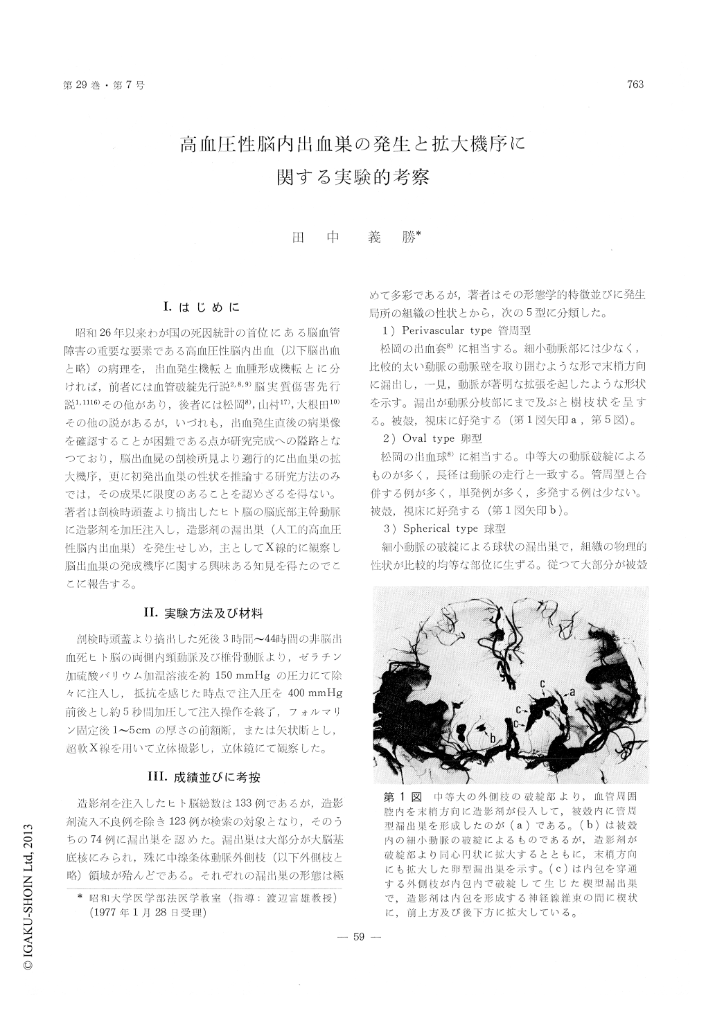
昭和26年以来わが国の死因統計の首位にある脳血管障害の重要な要素である高血圧性脳内出血(以下脳出血と略)の病理を,出血発生機転と血腫形成機転とに分ければ,前者には血管破綻先行説2,8,9)脳実質傷害先行説1,1116)その他があり,後者には松岡8)山村17),大根田10)その他の説があるが,いづれも,出血発生直後の病巣像を確認することが困難である点が研究完成への隘路となつており,脳出血屍の剖検所見より遡行的に出血巣の拡大機序,更に初発出血巣の性状を推論する研究方法のみでは,その成果に限度のあることを認めざるを得ない。著者は剖検時頭蓋より摘出したヒト脳の脳底部主幹動脈に造影剤を加圧注入し,造影剤の漏出巣(人工的高血圧性脳内出血巣)を発生せしめ,主としてX線的に観察し脳出血巣の発成磯序に関する興味ある知見を得たのでここに報告する。
For the purpose of clarifying the feature of the initial focus and the mode of growth at hyper-tensive intracerebral hemorrhage, the author per-formed the following experiment.
A gelatinized barium sulfate solution was injected with pressure into the vertebral artery and into the internal carotid arteries of 133 samples of human brains excised from skulls at the time of autopsy. Thus, hypertensive intracerebral hemorrhages were produced by barium artificially, and observed many initial foci and the extending features of leaked barium at bursting points in stereoscopically, useing the super soft X-ray.
These foci were classified in five groups analyzing its morphological characteristics of foci and physical property of adjacent tissue.
1) Perivascular type.
Leaked barium extends into Virchow-Robin's space of moderate arteries in size.
2) Oval type.
The major axis of leaked barium extends along the burst artery as well as the minor axis expands into the concentric circle from the bursting point of artery.
3) Spherical type.
Occurs in ganglia and not in white matter.
4) Wedging type.
This type shows a characteristic slender shape where one end is pointed and the other is spherical occurring in white matter especially in the internal capsule, mostly may be accompanied with the peri-vascular type.
5) Semilunar type.
Occurs in the external capsule closely connected to the putamen, therefore, the inside shows concave and the outside covex, and reveals on the both frontal and horizontal sections of the brain as the semilunar barium depot.
On the basis of the above facts, the author divided the growthing processes of bleeding focus into four steps.
A) First step. Mechanical growth
This is the step that the extending process is continued until keep a balance the pressure of leaking blood at the bursting point of artery with the mechanical resistance of adjacent tissue.
B) Second step. Peripheral growth.
The lesion is extended as a result of after bleeding due to circulatory disturbance in the peripheral area.
C) Third step. Neighboring growth.
The irregularly shaped hematoma changes into flat shape, and at the same time, gradually increases its volume.
D) Fourth step. Heterotopic growth.
As a result of oppression due to advanced intra-cranial pressure bleeding occurs in the area which is controlled by main arteries and veins, and they promote the growth of the already established hematoma and at times new bleeding in a location far from the above-mentioned hematoma.
The leaked foci as shown in the above 1) to 5) types, show simillar conditions of the initial bleeding foci of living body, and the author believe that the bleeding focus of a living body develops into massive hematoma through the four steps as shown the above-mentioned A) to D).
According to autopsys, we can find some examples which the extension of bleeding foci were stoped in early stage and the lesions had been modified to a cyst or cerebral softening as a result of healing process, and many other examples which the four steps had been progressed rapidly called as sudden death clinically. Both the former and the latter are contained with in author's classification and division.
Copyright © 1977, Igaku-Shoin Ltd. All rights reserved.