Japanese
English
- 有料閲覧
- Abstract 文献概要
- 1ページ目 Look Inside
I.はじめに
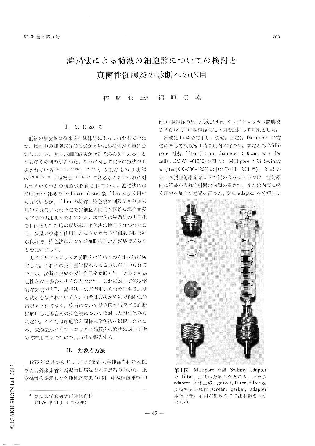
髄液の細胞診は従来遠心塗沫法によって行われていたが,操作中の細胞成分の損失が多いため検体が多量に必要なことや,著しい細胞破壊が診断に影響を与えることなど多くの問題があつた。これに対して種々の方法が工夫されている1,5,9,10,13〜19)。このうち主なものは沈澱法5,9,10,16,19)と濾過法1,14,15,17)であるがこのいづれに対してもいくつかの問題が指摘されている。濾過法にはMillipore社製のcelluiose-piastic製filterが多く用いられているが,filterの材質上染色法に制限があり従来用いられていた染色法では細胞の同定が困難な場合が多く本法の実用化が遅れている。著者らは濾過法の実用化を目的として細胞の収集率と染色法の検討を行つたところ,少量の検体を使用したにもかかわらず細胞の収集率が良好で,染色法によつては細胞の同定が容易であることを見い出した。
更にクリプトコッカス髄膜炎の診断への応用を特に検討した。これには従来墨汁標本による方法が用いられていたが,診断に熟練を要し発見率が低く4),培養でも偽陰性となる場合が少くなかつた6)。これに対して免疫学的な方法2,3,6,7),濾過法8)などが用いられ診断率を上げる試みもなされているが,前者は方法が繁雑で偽陽性の出現もまれでなく,後者については真菌性髄膜炎の診断に応用した場合その染色法について検討した報告はみられない。ここでは細胞診と同様に染色法を選択したところ,濾過法がクリプトコッカス髄膜炎の診断に対して極めて有用であつたので合わせて報告する。
Several techniques for cerebrospinal fluid (CSF)cytomorphology have been used, including centri-fuge-smear method, sedimentation method, filtrationmethod and others.
The centrifuge-smear method often results insevere distortion of cells and it requires a largevolume of CSF as a great number of cells maybe lost. The sedimentation method recently de-veloped has some benefits for identification of cellsin CSF, but requires a large volume of CSF as inthe centrifuge-smear method.
The membrane filter (the product of Milliporecompany) has been widely used to collect cells forCSF cytomorphology because the loss of cells isavoided in this method. It was revealed that thismethod using only 1.0ml of CSF enabled to collect40 to 80 per cent of the number of the cellsestimated in the counting chamber of Fuchs-Rosenthal. This fact shows that the examinationfor CSF cytomorphology is available with a littlevolume of CSF.
In the filtration method, some stains do not worksatisfactorily. The hematoxylin or the hematoxylin-eosin stain has been used in this method but thesestains have a disadvantage to identify the cells inCSF cytomorphology because of poorly stainedcytoplasm. The modified Gomori's trichrome staincould be satisfactorily applied to the filtrationmethod and adequately showed the morphologicfeatures of cells. The most suitable pH of Gomori'ssolution was 12.
The cytological diagnosis of cryptococcal menin-gitis may be difficult because of the frequent falsenegative data in an Indian ink preparation or ina culture for Cryptococcus neoformans. It was re-ported that the filtration method was available forthe diagnosis. In our experience, even by thismethod, it was difficult to identify cryptococciwhen the CSF contained insufficient number oforganisms. We could easily find even a feworganisms in the whole microscopic field by theAlcian blue stain or PAS stain because they werestrongly stained.
It is concluded that the filtration method isavailable for CSF cytomorphology and the diagnosisof cryptococcal menigitis if the suitable stains areselected for each case and the technique can beaccomplished quickly at the bed side, using onlya little volume of CSF.

Copyright © 1977, Igaku-Shoin Ltd. All rights reserved.


