Japanese
English
- 有料閲覧
- Abstract 文献概要
- 1ページ目 Look Inside
I.はじめに
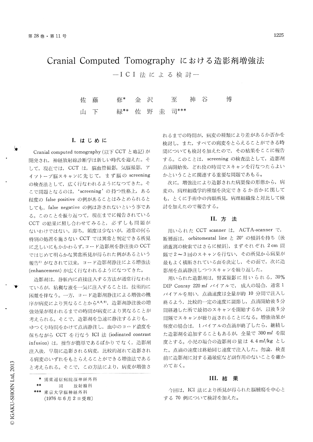
Cranial computed tomography (以下CCTと略記)が開発され,神経放射線診断学は新しい時代を迎えた。そして,現在では,CCTは,脳血管撮影,気脳撮影,アイソトープ脳スキャンに先じて,まず脳のscreeningの検査法として,広く行なわれるようになつてきた。そこで問題となるのは,‘screening’の持つ性格上,ある程度のfalse positiveの例があることはみとめられるとしても,false negativeの例は許されないという事である。このことを振り返つて,現在までに報告されているCCTの結果に照し合わせてみると,必ずしも問題がないわけではない。即ち,頻度は少ないが,通常の何ら特別の処置を施さないCCTでは異常と判定できる所見に乏しいにもかかわらず,ヨード造影剤を静注後のCCTではじめて明らかな異常所見が得られた例があるという報告1)がなされて以来,ヨード造影剤静注による増強法(enhancement)が広く行なわれるようになつてきた。
造影剤は,静脈内に直接注入する方法が通常行なわれているが,粘稠な液を一気に注入することは,技術的に困難を伴なう。一方,ヨード造影剤静注による増強の機序が病変により異なることから4,8,9),造影剤静注後の増強効果が現われるまでの時間が病変により異なることが考えられる。そこで,造影剤を急速に静注するよりも,ゆつくり時間をかけて点滴静注し,血中のヨード濃度を保ちながらCCTを行なうICI法(iodinated contrastinfusion)は,操作が簡単であるばかりでなく,造影剤注入後,早期に造影される病変,比較的遅れて造影される病変のいずれをもとらえることができる増強法であると考えられる。そこで,この方法により,病変が増強されるまでの時間が,病変の種類により差があるか否かを検討し,また,すべての病変をとらえることができる時間についても検討を加えたので,その結果をここに報告する。このことは,screeningの検査法として,造影剤点滴開始後,どれ位の時間でスキャンを行なつたらよいかということに関連する重要な問題であもる。
次に,増強法により造影された病巣像の形態から,病変の,病理組織学的種類を決定できるか否かに関しても,とくに手術中の肉眼所見,病理組織像と対比して検討を加えたので報告する。
The effect of infusion of iodinated contrast material on the enhancement of the computed tomography were studied in several kinds of the intracranial lesions.
Especially, relationship between the degree of the enhancement and the time interval after the infusion was examined.
It was found that the appearance of the en-hancement effect were different according to the pathological types of the lesions. There were also time differences even in the same lesion before the full size of the lesions was demonstrated.
The pathological type of the lesions could be suggested from the enhanced figures of the lesions, but the diagnosis would be more accurate in con-sideration of the localization of the lesions.
It was thus concluded that this method was useful for the detecttion of the precise localization and the size of the lesions which were not so clearly demonstrated in conventional cranial computed tomography.

Copyright © 1976, Igaku-Shoin Ltd. All rights reserved.


