Japanese
English
- 有料閲覧
- Abstract 文献概要
- 1ページ目 Look Inside
I.はじめに
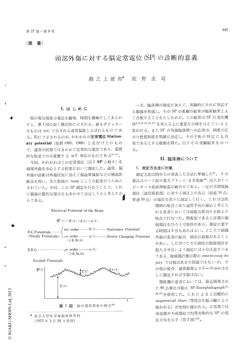
脳の電気現象は電位を縦軸,時間を横軸にしてあらわすと,第1図の如く模式的に示される。最もポピュラーなものはe (t)で示される通常脳波とよばれるものである。E (t)で示されるのが,われわれが定常電位Station—ary potential (佐野1965,1966)と名付けたのもので,通常の状態ではきわめて定常的な電位であり,器質的な疾患でのみ変動するmV単位のものである9〜11)。
今回,われわれはこの定常電位(以下SPと略す)を頭部外傷を中心とする疾患において測定した。通常,脳外傷の診断は外傷状況に加えて脳血管撮影などの補助診断法を用い,主に損傷のmassとしての結果がうんぬんされていた。今回,このSP測定を行なうことで,これに損傷の質的な部分をもあわせて決定しうると考えたからである。
一方,臨床例の測定に加えて,実験的にそれに対応する損傷を作成し,そのSPの変動の結果が臨床結果とよく合致することをたしかめた。この結果はSPの発生機序5,6,9〜15,23,27を考える上に重要な示唆を与えていると思われる。またSPの外傷臨床例への応用は,病変の広がり性質程度を明確に決定し,その予後の判定にも有用であるとする根拠を得た。以下その実験結果をのべる。
An analysis of brain injury was attemted clinical-ly and experimentally by recording stationarypotential (SP) which is most stable component ofthe three electrical phenomena of the brain (Fig. 1).SP value is considered to vary only when thereare organic lesions or severe metabolic change inthe cerebral hemisphere and also epileptic seizures.Conversely, the organic cerebral disease could bediagnosed by monitoring SP change.
Clinically, the SP changes have been measuredon 103 head injured patients. If an organic lesionis located in the cerebral hemisphere, the SP showsvarious change according to the location and natureof the lesion (Table 1).
Recording method was shown in Fig. 8. Givingan outline, the combination of calomel half-cellelectrodes, salt brigde, high impedance DC ampli-fires (input impedence is ten thousands MG ormore) was adopted and usually SP values weremeasured at least at thirty points on the scalp.
Positive SP changes are usually seen in the casesof mild cortical compression and subcortical lesions.Clinically, these lesions are equivalent to acuteepidural hematoma, acute subdural hematoma with-out contusion, chronic subdural hematoma, simpledepressed fracture and intracerebral hematomawithout cortical damage respectively.
Whereas, almost all of the cortical lesions or ex-tensive brain lesions show negative changes of SP.Such cases are cerebral contusion, intracerebralhematoma with vascular lesion, depressed fracturewith cortical damage and traumatic epilepsy (Fig. 7).
In these clinical data, we could observe fairyaccurate correlation between the positive or negativeSP change and the prognosis of the patients. Thatis, the group with positive SP change shows atendency to have good prognosis in mortality andmorbidity and contraversely, the prognosis ofnegative group, especially cerebral contusion, isnot good generally.
Subsequently, to make support these data, 1) ex-perimental cortical compression with clot insertedinto subdural and extradural space, 2) subcorticallesion by artificial intracerebral hematoma, 3) cere-bral contusion by mechanical destruction in catswere investigated, recording respective SP changes.The result show the same tendency in accordancewith the clinical data (Fig. 9, 10).
In another experimental study, we tried to clarifythe possibility of SP change in cerebral concussionand observed that the SP of concussed animal wasrecorded as negative almost inevitably (Fig. 11, 12).
We are of opinion that SP recording especiallyin acute stage of head injury is one of the mostuseful diagnostic method because SP is very simpleto measure without any complication and we caneasily and precisely predict the nature and extensionof the lesion from the distribution of SP change.

Copyright © 1975, Igaku-Shoin Ltd. All rights reserved.


