Japanese
English
- 有料閲覧
- Abstract 文献概要
- 1ページ目 Look Inside
はじめに
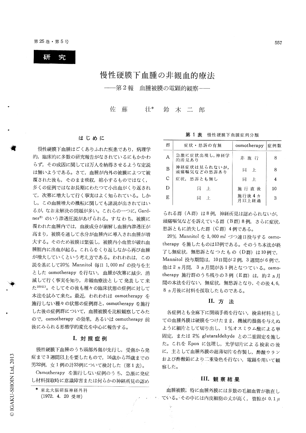
慢性硬膜下血腫はごくありふれた疾患であり,病理学的,臨床的に多数の研究報告がなされているにもかかわらず,その成因に関しては万人を納得させるような定説は無いようである。さて,血腫が内外の被膜によつて被覆された後も,そのまま吸収,縮小するものではなく,多くの症例ではなお長期にわたつて小出血がくり返されて,次第に増大して行く事実はよく知られている。しかし,この血腫増大の機転に関しても諸説が出されてはいるが,なお未解決の問題が多い。これらの一つに,Gard—ner3)のいう滲透圧説があげられる。すなわち,被膜に覆われた血腫内では,血液成分が崩解し血腫内滲透圧が高まり,被膜を通して水分が血腫内に導入され血腫が増大する。そのため被膜は緊張し,被膜内小血管が破れ血腫腔内に出血が起る。これらをくり返しながら再び血腫が増大していくという考え方である。われわれは,この説を基にして20%Mannitol毎日1,000mlの投与を主としたosmotherapyを行ない,血腫が次第に減少,消滅して行く事実を知り,非観血療法として発表して来た10)11)。そしてその後も種々の臨床状態の症例に対して本法を試みて来た。最近,われわれはosmotherapyを施行しない種々の状態の症例群と,osmotherapyを施行した後の症例群について,血腫被膜を比較観察してみたので,osmotherapyの効果,あるいはosmotherapy前後にみられる形態学的変化を中心に報告する。
Thirty-three cases of chronic subdural hematoma were classified into five groups as follows : A) cases with neurological deficits or disturbance of cons-ciousness, B) cases without any neurological signs but with the complaints of headache and nausea, C) cases without any signs or symptoms, D) cases without any signs or symptoms after the osmo-therapy for 10-21 days, E) cases without any signs or symptoms four months or more after the osmo-therapy. In the osmotherapy we used intravenous drips of 20% Mannitol, 1,000 ml daily. Specimens of the capsule of these groups were obtained on surgery and investigated by light and electron microscope to know ultrastructural changes of it before and after the osmotherapy.
In the capsule of group A, there were many growing blood capillaries which possessed endo-thelial cytoplasmic processes or pseudoped like protrusions. The protrusion was less in group D than group A, B, C and it was further less in group E. Pinocytotic vesicles of the endothelial cellswere few in group A, and increased in number as clinical symptomes were improved. Endothelial fenestrations or open gaps between adjacent cells were often found in group A. They decreased in group B and C, and could not be found in group D and E. Sometimes degenerated endothelial cells were observed among normal endothelial cells of growing capillaries. They appeared more in group A than group B, C and were no more found in group D and E. As a result, in cases with neuro-logical deficits, permiability of vessels increased and endothelial degenerations of its vessels were revealed. These morphological changes were res-tored to the normal condition by the osmotherapy.
Abundant collagen fibrils and fibroblasts were seen in the hematoma capsule. Smooth surfaced endoplasmic reticula, which contained collagen fibrils, were occasionally observed in the cytoplasm of fibroblasts. It seemed one of many types of collagen formation. The osmotherapy promoted the formation of collagen fibrils in the hematoma capsule, because the reticulum was found more frequently in group D.

Copyright © 1973, Igaku-Shoin Ltd. All rights reserved.


