Japanese
English
- 有料閲覧
- Abstract 文献概要
- 1ページ目 Look Inside
I.はじめに
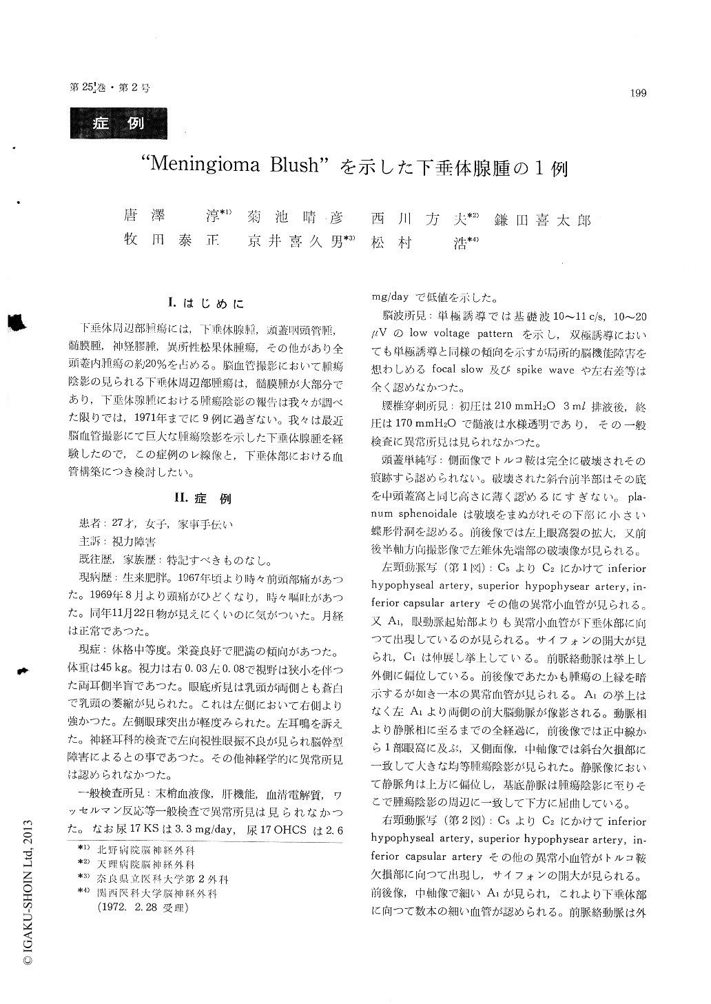
下垂体周辺部腫瘍には,下垂体腺腫,頭蓋咽頭管腫,髄膜腫,神経膠腫,異所性松果体腫瘍,その他があり全頭蓋内腫瘍の約20%を占める。脳血管撮影において腫瘍陰影の見られる下垂体周辺部腫瘍は,髄膜腫が大部分であり,下垂体腺腫における腫瘍陰影の報告は我々が調べた限りでは,1971年までに9例に過ぎない。我々は最近脳血管撮影にて巨大な腫瘍陰影を示した下垂体腺腫を経験したので,この症例のレ線像と,下垂体部における血管構築につき検討したい。
The patient was 27 year-old and female. She had sometimes suffered from frontalgia since 1967. She had had severe headache since August, 1969 and occasional vomiting. As she realized distur-bance of vision on November 22, 1969, she was admitted to our hospital on January 8, 1970.
The constitution was middle. The visual acuity of the right eye was 0.03 and one the left eye was O. 08. The visual field was bitemporal hernia-nopsia accompanied by contraction. In the findings of optic fundi, optic atrophy was admitted in the bilateral eyes. Slight exophthalmus was recognized in the left eye. She complained tinnitus in the left ear. Other abnormalities were not admitted in the cranial nerves. Cerebral and cerebellar signs and pathological reflex were not recognized either.
Skull radiography showed total destruction of the sella turcica and the same level of the anterior part of clivus as the middle fossa. The anterio-posterior view indicated enlargement of the superior orbital fissura and the anterio-posterior half axial view showed destruction of the left pyramidal appex.
The left carotid angiography revealed the inferior hypophyseal artery, superior hypophyseal artery, inferior capsular artery and other abnormal small vessels. Abnormal small vessels were also visualized from the part of Ai, proximal part of ophthalmic artery to the tumor. Tumor stain was evenly recognized from the median line to part of the left orbit in the antero-posterior view and from the arterial phase to the venous phase corres-ponding to the defective part of clivus in the lateral view.
The right carotid angiography visualized the inferior hypophyseal artery, superior hypophyseal artery, inferior capsular artery and other abnormal small vessels towards the part of sella turcica. No tumor stain was recognized. Vertebral angio-graphy showed that a basilar artery remarkably stretched and was displaced posteriorly. The left posterior cerebral artery and the left superior cerebellar artery indicated upward displacement at their proximal parts.
Pneumoencephalotomography visualized tumor with a width of over 3 cm from the median line to both sides. The tumor shadow of the right was smooth on the surface but one of the left was uneven.
From the above neuroradiological findings, tumor was found to have orbital, hypothalamic, temporal and posterior subtentorial extension. The operation was performed by right-frontolateral sphenoidal approach on January 19, 1970. It was, however, limited to the partial removal due to the difficulty of controlling the bleeding. The pathological di-agnosis of the specimen at that time was chromo-phobe adenoma. There have been 9 cases of pi-tuitary adenoma complicated with tumor stain reported by Ecker and Riemenschneider, Kricheff and Schotland, Lehrer and Richardson, Cophignon et al, Davis by 1971. Our case seems to be the 10th reported case. Judging from neuroradiological findings of our patient's tumor, the left side was assumed to be more malignant than the right side. The patient was discharged after the radiation therapy and is now engaged in domestic chores.

Copyright © 1973, Igaku-Shoin Ltd. All rights reserved.


