Japanese
English
- 有料閲覧
- Abstract 文献概要
- 1ページ目 Look Inside
I.緒言
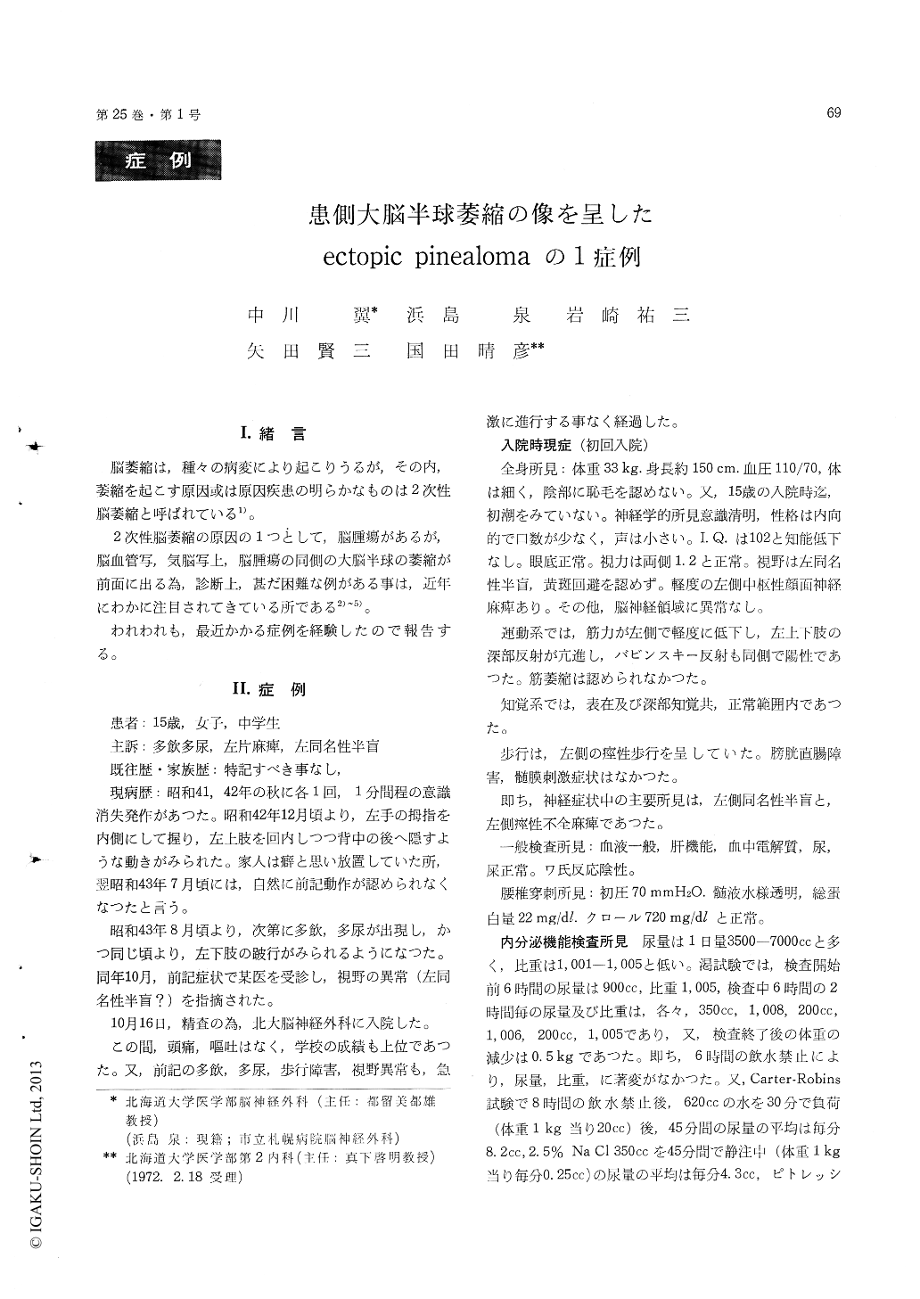
脳萎縮は,種々の病変により起こりうるが,その内,萎縮を起こす原因或は原因疾患の明らかなものは2次性脳萎縮と呼ばれている1)。
2次性脳萎縮の原因の1つとして,脳腫瘍があるが,脳血管写,気脳写上,脳腫瘍の同側の大脳半球の萎縮が前面に出る為,診断上,甚だ困難な例がある事は,近年にわかに注目されてきている所である2)〜5)。
Although dilatation of the ventricular system is commonly ohserved in case with brain tumors, especially in those which is related to cerebrospinal fluid passway, it is rare to observe secondary atrophy of the ipsilateral cerebral hemisphere before removal of the neoplasm, and also without increased intra-cranial pressure. The authors found only five such cases in the literature, all of which were the cases with ectopic pinealoma.
The patient reported here is a 15-year old Japanese female who was admitted to Hokkaido University Hospital on Oct. 16, 1968 with chief complaints of difficulty in walking, impaired visual fields, poly-dipsia and polyuria. These symptoms have been first noticed approximately two years prior to admis-sion and did not change much till the time of the admission. Examination on admission revealed left homonymous hemianopia without macular spairing, spastic hemiparesis on the left side and diabetes incipidus. There were no signs or symptoms to indicate increased intracranial pressure. Cerebro-spinal fluid pressure measured by lumbar puncture was 70 mm H2O, and it contained 22 mg/dl of total protein. Plain skull X-ray revealed no abnormalities, and no calcification was seen in the region of the pineal body. Carotid angiogram showed displace-ment of the anterior cerebral artery toward right side (fig. 1). Pneumoencephalogram with lamino-graphic studies showed dilatation of the anterior part of the right lateral ventricle and enlargement of the cerebral sulci on the same side (fig. 2-3). Because of these findings, without establishing de-finitive diagnosis, the patient was discharged on Feb. 5, 1969, to be followed by out-patient department.
The patient was readmitted on June 9, 1969, be-cause of progression of the left hemiparesis and of disturbance of consciousness. Lumbar puncture on this admission showed initial pressure of 170 mm H2O with total protein of 147 mg/dl. Pneumoence-phalogram was also repeated and it showed further advancement of the dilatation of the right lateral ventricle and widening of the cerebral sulci on the same side (fig. 4).
Exploratory craniotomy was performed on Aug. 27,1969. The entire surface of the right lateral ventricle was covered with grayish-white material of which histological examination turned out to be a typical two-cell pattern pinealoma (fig. 5). Radiation therapy using CO60 was started following to the surgery, but the patient took downhill course and expired on Sept. 16, 1969. On postmortem ex-amination, despite the fact that the neoplastic tissue was diffusely invading widely including the basal ganglia, the internal capsule, the thalamus and the hypothalamus on the right side, the right optic tract, and entire surface of the ventricularsystem, the right cerebral hemisphere showed dif-fuse atrophy which was most marked in the frontal lobe. It was failed to find any neoplastic cells in the pineal body itself.
It is quite interesting that this type of infiltrat-ing neoplasm could cause diffuse atrophy of the cerebral hemisphere before it produces cerebralswelling or blockage of the cerebrospinal fluid pass-way. It is also interesting that despite the fact this type of neoplasm tends to spread along the ventricular wall, the protein content in the cerebro-spinal fluid does not elevate in the early stage.

Copyright © 1973, Igaku-Shoin Ltd. All rights reserved.


