Japanese
English
- 有料閲覧
- Abstract 文献概要
- 1ページ目 Look Inside
はじめに
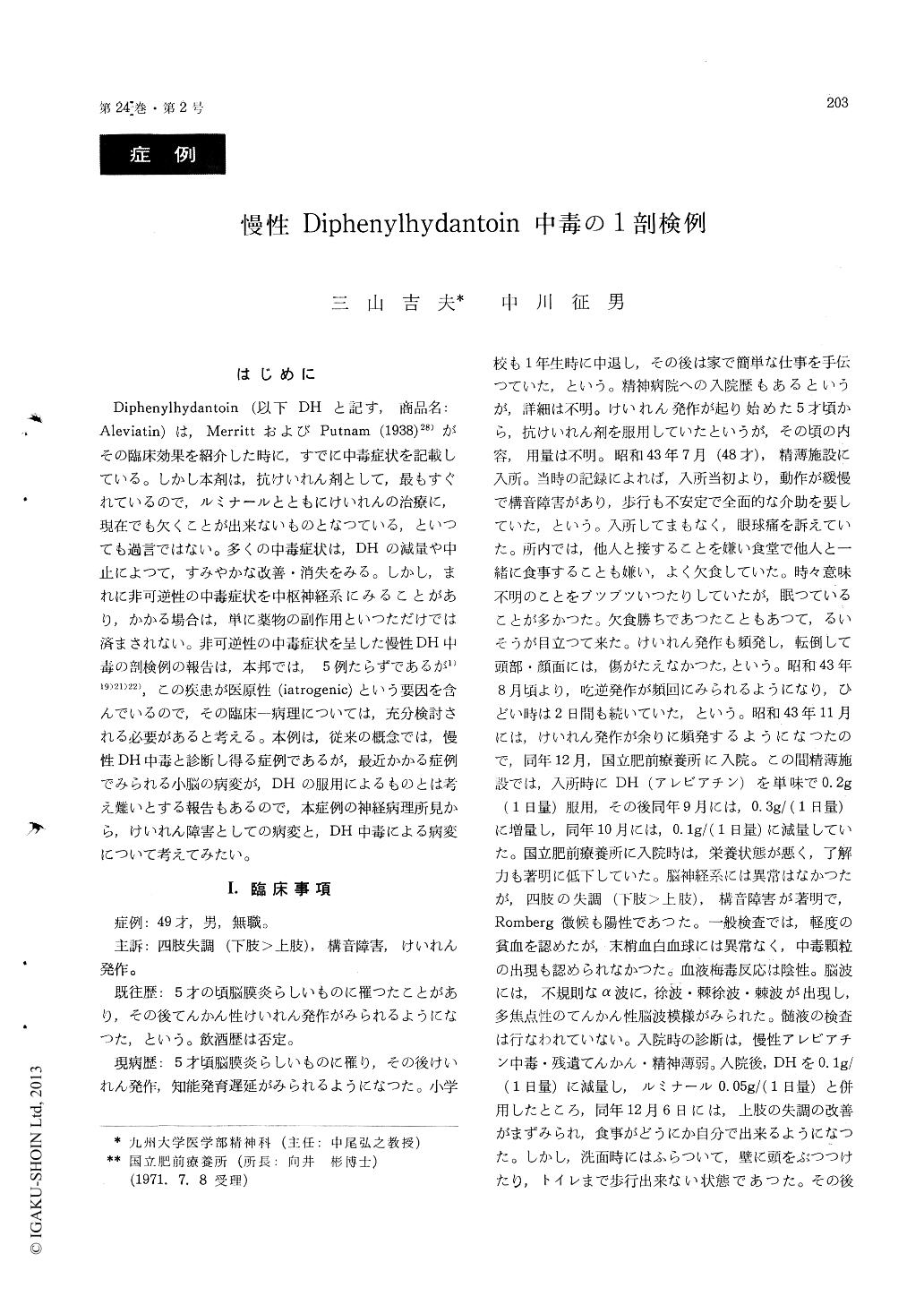
Diphenylhydantoin (以下DHと記す,商品名:Aleviatin)は,MerrittおよびPutnam (1938)28)がその臨床効果を紹介した時に,すでに中毒症状を記載している。しかし本剤は,抗けいれん剤として,最もすぐれているので,ルミナールとともにけいれんの治療に,現在でも欠くことが出来ないものとなつている,といつても過言ではない。多くの中毒症状は,DHの減量や中止によつて,すみやかな改善・消失をみる。しかし,まれに非可逆性の中毒症状を中枢神経系にみることがあり,かかる場合は,単に薬物の副作用といつただけでは済まされない。非可逆性の中毒症状を呈した慢性DH中毒の剖検例の報告は,本邦では,5例たらずであるが1)19)21)22),この疾患が医原性(iatrogenic)という要因を含んでいるので,その臨床—病理については,充分検討される必要があると考える。本例は,従来の概念では,慢性DH中毒と診断し得る症例であるが,最近かかる症例でみられる小脳の病変が,DHの服用によるものとは考え難いとする報告もあるので,本症例の神経病理所見から,けいれん障害としての病変と,DH中毒による病変について考えてみたい。
Case : A 49-year-old male had suffered from mental retardation and general convulsions that began at age 5 as residual syndrome of meningitis at the same age. He had been treated with anticonvulsants for about 45 years. About 10 months prior to his death, he had developed cerebellar ataxia with 0. 2- O. 3 g per day of diphenylhydantoin (Aleviatin). He complained of weakness and inability to eat solid food. His gait became staggering, and he lost balance very easily and fell frequently. These cere-bellar syndrome were brought to incomplete remis-sion by reduction of the diphenylhydantoin dose. But, his epileptic seizures were frequent. He died of acute bronchopneumonia.
Postmortem findings : The general autopsy reveal-ed no remarkable findings except for bilateral bron-chopneumonia.
Gross examination of the brain : The brain weighed 1250 g, which showed two small traumatic scars on the left frontal and parietal lobe but no ulegyria cerebri. The folia of the cerebellar hemisphere were mildly atrophic without any localized lesion.
Histological examination : The most prominent pathology was cerebellar cortical change consisting of an almost complete disappearance of Purkinje cells from the bilateral ventral surfaces in the cere-bellar hemisphere. Here and there a pyknotic dark stained or a ghost Purkinje cell could be detected and there was a mild fibrillary gliosis in the me-dullary lamina of the folia.
On the dorsal surface, Purkinje cells were normally preserved. Where Purkinje cells had disappeared, Bergmann's glia were markedly increased in number and size. The granular layer showed no markedchange. In specimens impregnated with silber, axonal changes of Purkinje cell could be detected in the granular layer. The empty basket structures were generally well preserved.
The bilateral Ammon's horns revealed fibrillary gliosis with almost complete loss of nerve cells in end-plate and Sommer's sector.
The remainder of the brain revealed diffuse marginal gliosis of a moderate degree.
These findings bore much resemblance to those of autopsy cases and animal experiments with di-phenylhydantoin intoxication in the literature.
In the production of these lesions, three factors seem to have pathogenic effect : a) individual sensi-tivity to diphenylhydantoin, b) diphenylhydantoin administration over a long period, and c) epileptic seizures.

Copyright © 1972, Igaku-Shoin Ltd. All rights reserved.


