Japanese
English
- 有料閲覧
- Abstract 文献概要
- 1ページ目 Look Inside
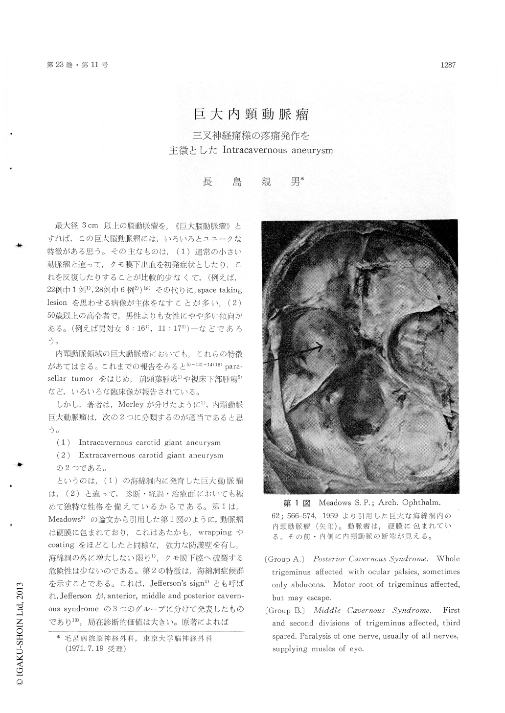
最大径3cm以上の脳動脈瘤を,《巨大脳動脈瘤》とすれば,この巨大脳動脈瘤には,いろいろとユニークな特徴がある思う。その主なものは,(1)通常の小さい動脈瘤と違って,クモ膜下出血を初発症状としたり,これを反復したりすることが比較的少なくて,(例えば,22例中1例1),281例中6例2))18)その代りに,space takinglesionを思わせる病像が主体をなすことが多い,(2)50歳以上の高令者で,男性よりも女性にやや多い傾向がある。(例えば男対女6:161),11:172))—などであろう。
内頸動脈領域の巨大動脈瘤においても,これらの特徴があてはまる。これまでの報告をみると5)〜12)〜14)18)para—sellar tumorをはじめ,前頭葉腫瘍1)や視床下部腫瘍5)など,いろいろな臨床像が報告されている。
This paper presents a case of the intracavernous carotid aneurysm of extralarge globoid type with 5cm × 4. 5cm × 5cm on angiographic measurements which, of interesting to note, had sudden onset of the tic douloureux persisted for 2 months prior to admission.
The patient is 63-year-old-female who developed lockjaw. Three days later it was followed by in-tense stabs of pain over the right half of the face 30 minutes to one hour in duration and recurring as often as several times daily. The pain started from the right retroauricular region spreading tothe right forehead, eye, chinn and lower jaw, which forced the patient to compress tightly over right half of the face with her right hand. In inter-paroxysmal period, the pain frequently focused into the right side of the nose and corner of the mouth. No definite trigger area was noted. Repetition of the attacks persisted for two months, then gradual-ly lessend but she had been rarely free from tingl-ing of the face. On admission her blood pressure was 130/90 mmHg.
There was hypesthesia over the area of the first, second and third branches of the right trigeminal nerve, trismus of 2-finger-breadth, loss of taste over the anterior 2/3 of right half of the tongue, optic atrophy more on the right eye, the oculo-motor, trochler and abducens paresis of the right eye. Wasserman reaction was negative.
Tentative diagnosis was tumor invading the right middle fossa. Basal skull X-ray showed complete erosion of the petrous part of the temporal bone with probable intramural calcification outlining the mass (Fig. 2). Lateral skull film showed curvilinear calcifications above and behind the sella turtica with rarefaction of the posterior clinoid process. (Fig. 5). Angiography, however, revealed a huge intracavernous globoid aneurysm (Fig. 3, 4). Uni-lateral enlargement of the right superior fissurewith erosion of the greater and lesser wings of the sphenoid was also noted (Fig. 5).
First operation : Adequate cross-filling demonst-rated by angiography prompted the author to ligate the right common carotid artery to relieve the pain. It resulted immediate loss of facial pain and gradual improvement of taste on the tongue and visual acuity of both eyes.
However, follow-up angiogram, done at post-operative 15 months through the retrograde injec-tion into the brachial artery still showed the aneu-rysm of nearly 1/2 of pre-operative size (Fig. 6).
This retrograde vertebral arteriogram showed a new anastomosis between the right vertebral and ipsilateral external carotid artery ; the ipsilateral internal carotid and the aneurysm were filled maily by this pathway. The dilated posterior communi-cating artery filling the aneurysm was also visuali-zed.
Second operation ; Trapping of the aneurysm by clipping the intracranial internal carotid and by ligating the cervical internal carotid artery was carried out with uneventuful post-operative course. The patient is free of symptom except for diplopia and mild hypesthesia of right half of the face.

Copyright © 1971, Igaku-Shoin Ltd. All rights reserved.


