Japanese
English
- 有料閲覧
- Abstract 文献概要
- 1ページ目 Look Inside
I.緒言
脳循環を論ずるにさいし,全体的な頭蓋内圧亢進が問題となっていたが,近来は脳の特定部位における循環障害または血管性障害注目されつつある。Lindenberg1)は脳圧亢進を起こすいかなる病的状態においても動脈圧迫は起こり,その支配領域に病変を生ずるという血管圧迫説で淡蒼球その他脳の局在性壊死の好発部位の解析を行なっており,われわれの教室の小林2)は,脳浮腫と動脈圧迫効果特に基底核における選択的病変の成立機序について報告している。著者は,今回小林の研究をさらに脳ヘルニアの分類にまでさ発展せた。すなわち,頭蓋内血腫・脳腫瘍や脳浮腫など脳の占拠性障害(space—occupying intracranial lesion)として,脳実質の一部が頭蓋内の隔壁である天幕や大脳鎌,頭蓋底の境界であるsphenoid ridge,開口部である大後頭孔などを越えて他の区画にはみ出すことを脳ヘルニアという。この脳ヘルニアはMeyer3)により初めて記載されて以来,多くの人々の報告があり,Meyer3)は3群に,Ecker4)は5群に,Azambujaら5)と石井6)は4群に分類している。従来の分類では脳実質の一部が頭蓋内のどの間隙からはみ出すかということと,脳実質のどの部分がはみ出すかということが混然としているものが多い。脳ヘルニアの分類は,たんに形態の変化を表現するにとどまらず,脳ヘルニアによって招来する臨床症状の識別にまで発展さすべきであり,また,種々の原因による頭蓋内圧亢進の救急治療の根本は,脳ヘルニアの解除にあるといつても過言ではない。脳実質がどの間隙からはみ出すかということよりも,脳実質のどの部分がはみ出すかが重要であり,しかも前者と後者は必ずしも一致しないことに着目した著者は,剖検脳について脳ヘルニアの実態を把握するとともに,5群に分類したのでここに報告する。
Cerebral herniation refers to protrusion of a por tion of the brain across an intracranial septum (tentorium cerebelli, falx cerebri) or boney border (sphenoid ridge), or into the foramen magnum be-cause of increased intracranial pressure, which may be caused by a hematoma or tumor, or by cerebral swelling alone. Generalized cerebral swelling may be secondary to a jarring type of trauma, such as repeated blows or punches to the head.
It is also important to know which portion of theswollen brain has herniated.
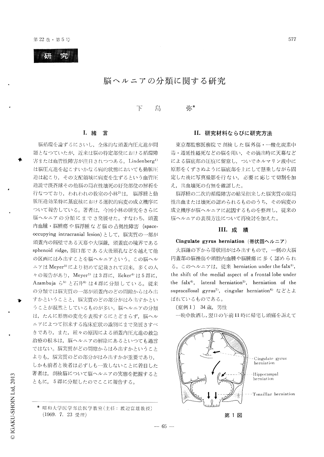
(1) Cingulate gyrus herniation:
Protrusion to the opposite side of the cingulate gyrus beneath the lower edge of the falx cerebri.
(2) Orbital gyrus herniation:
This portion of the base of the frontal lobe, after filling the Sylvian fissure, extends over the lesser wing of the sphenoid bone into the middle cranial fossa.
(3) Anterior perforated substance herniation :
As the result of the cerebral swelling, in addition to a downward pressure on the brain stem, there may be herniation of the anterior perforated sub-stance and adjacent part of the olfactory tracts against the ridge of the sphenoid and edge of the tentorium. Pressure on the anterior and middle striate arteries and venous structures may result in a hemorrhagic necrosis corresponding to the distribu-tion of these vessels.
(4) Hippocampal herniation :
The uncus and hippocampus herniate beneath the edge of the tentorium cerebelli.
(5) Tonsillar herniation :
The cerebellar tonsils herniate into the foramen magnum after expanding into the cisterna magnum.

Copyright © 1970, Igaku-Shoin Ltd. All rights reserved.


