Japanese
English
- 有料閲覧
- Abstract 文献概要
- 1ページ目 Look Inside
I.緒言
頭蓋内疾患において,視力低下と視野の欠損を主症状とする疾患は脳神経外科領域においてきわめて多い。
そのなかでもpituitary adenoma, craniopharyngio—ma, parasellar meningioma, optic nerve glioma, opticochiasmatic arachnoiditisおよびsacular aneu—rysmaなどがその代表的なものとされ,その診断方法および治療についてはすでに多くの報告がある。
A case of visual loss associated with fusiformenlargement of the intracranial portion of the in-ternal carotid artery of a 48 year old man was reported.
At the onset, he awoke morning and noted that felt right visual acuity. After two days, vision in the right eye was almost entirely gone. At this time he also complained of right frontal head pain. This lasted two weeks and gradually the vision improved, but objects appeared blurred. There were no other symptoms and the left eye was normal.
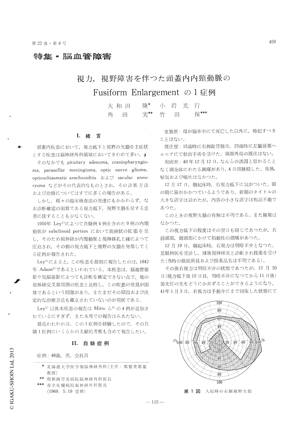
He was admitted in January, 1966. At this time his vision on the right side was such that he could barely count fingers at 15cm ; the inferior field was absent and macular portions were not remained (Fig. 1). The right disc was now pale but left was within normal limits. Blood pressure was 120/70. His blood picture, uninalysis, serum proteins and electrolytes were normal. The Wassermann and Kahn reactions of the blood was negative. The chest and skull films which included optic foramen views were negative. Lumbal puncture yielded clear, color-less spinal fluid under an initial pressure of 120 mm of water. The total protein content of the spinal fluid was 41 mg. per hundred cubic centimeters. An electroencephalogram was normal. Bilateral carotid arteriograms was performed and it was thought that a right carotid arteriogram indicated some enlargement of the intracranial portion of the internal carotid artery (Fig. 2, Fig. 3).
On January 26, 1966, a frontal craniotomy was carried out and the optic nerve was visualized on this side. It was compressed and thinned by a gross-ly enlarged carotid artery which displaced the nerve against the rim of the optic foramen (Fig. 4). Unroofing of the optic canal on the involved side was done. The past operative course was eventful : Both of the visual acuity and field was improved (Fig. 1, Fig. 4). When last seen, on May 20, 1966, his visual functions remained unchanged.
The authors reviewed the literatures and stressed upon the difficulty of the current diagnosis of the fusiform enlargement of the intracranial portion of the internal carotid artery before autopsy or opera-tion.
Copyright © 1970, Igaku-Shoin Ltd. All rights reserved.