Japanese
English
- 有料閲覧
- Abstract 文献概要
- 1ページ目 Look Inside
I.緒言
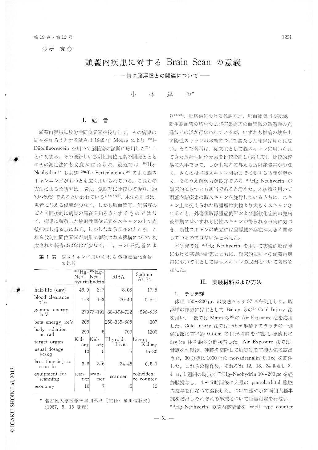
頭蓋内疾患に放射性同位元素を投与して,その病巣の局在を知ろうとする試みは1948年Mooreにより131I—Diiodfluoresceinを用いて脳腫瘍の診断に応用した20)ことに初まる。その後新しい放射性同位元素の開発とともにその測定法にも改良が重ねられ,最近では203Hg—Neohydrin4)および99mTc Pertechnetate10)による脳スキャンニングがもつとも広く用いられている。これらの方法による診断率は,脳波,気脳写に比較して優り,約70〜80%であるといわれている1)8)18)25)。本法の利点は,患者に与える侵襲が少なく,しかも脳血管写,気脳写のごとく間接的に病巣の局在を知ろうとするものではなく,病巣に蓄積した放射性同位元素をスキャンの上で直接把握し得る点にある。しかしながら現在のところ,これら放射性同位元素が病巣に蓄積される機構について検索された報告ははなはだ少なく,二,三の研究者により14)29),脳病巣における代謝亢進,脳血液関門の破壊,新生脳血管の増生および病巣周辺の血管壁の透過性の亢進などの説が行なわれているが,いずれも推論の域を出ず陽性スキャンの本態について論及した報告は見られない。そこで著者は,従来主として脳スキャンに用いられてきた放射性同位元素を比較検討し(第1表),比較的容易に入手できて,しかも患者に与える放射能障害が少なく,さらに投与後スキャン開始までに要する時間が短かく,そのうえ解像力が良好である203Hg-Neohydrinが臨床的にもつとも適当であると考えた。本核種を用いて頭蓋内諸疾患の脳スキャンを施行しているうちに,スキャン上に捉えられた脳腫瘍は実物より大きくスキャンされること,外傷後脳浮腫症例22)および脳軟化症例の発病後早期にはいずれも陽性スキャンが得られる事実に気づき,陽性スキャンの成立には脳浮腫の存在が大きく関与しているのではないかと考えた。
本研究では203Hg-Neohydrinを用いて実験的脳浮腫における某礎的研究とともに,臨床的に種々の頭蓋内疾患において主として陽性スキャンの成因について考察を加えた。
57 brain scans using 203Hg-Neohydrin were per-formed.
Experimental and clinical studies were made con-cerning to the relationship between positive scan and brain edema. Experimental studies:
Brain edema induced by cold injury and air ex-posure was made on one side of cerebral hemisphere in 57 adult rats and 203Hg-Neohydrin was given. It was found that the 2031-Ig-Neohydrin accumulation in rat's edematous hemisphere was 4.5 times more than that of the contralateral hemisphere, while that of RISA was 2 times and that of Hippurate showed no significant difference. This ratio of Neohydrin accumulation seemed to well correlate with the degree and extent of brain edema.
In 13 dogs with brain edema induced by extradural ballooning and cold injury, higher RI counts were obtained in edematous hemisphere than in the con-tralateral hemisphere.
By the macroautoradiogram using 203Hg-Neo-hydrin localization of the edema was clearly shown in dog's brain and microautoradiogram of edematous rat's brain demonstrated that the silver grain by 203Hg-Neohydrin was mainly found within dilated capillary vessels and supposedly in the intercellular space. Clinical studies:
The total diagnostic accuracy of brain scans in 49 patients was 71.4% and the accuracy for brain tumor, intracranial hematoma, head injury and cerebro-vascular disease were 83.3%, 92.3%, 70% and 50% respectively.
1) Brain Tumor: There was significant accumulation of "3Hg-Neohydrin not only in the tumor tissue but also in the edematous brain tissue adjacent to the tumor. So, the positive scan of brain tumor might often look larger than its actual size.
2) Intracranial Hematoma: This group consisted of 10 cases with subdural, 2 with epidural and one with intracerebral hematoma. In 8 cases of subdural hematoma the accumulation of 203Hg-Neohydrin in the hematoma was not higher than that of blood, and the highest accumulation was obtained from the capsule. So, the positive scan of subdural hematoma might show three layers: the semilunar " cold area ", the "linear positive" layer and the "diffuse positive" layer and the " diffuse positive " layer, each corres-ponding to the hematoma, the capsule and the edema-tous brain tissue due to compression by the hematoma.
3) Head Injury : Ten cases of head injury were con-sidered to be traumatic brain edema and had re-covered within two weeks or more without any neurological abnormality remained. Seven out of 10 cases were positive on initial scan (70%). Selecting the cases in which the time interval from injury to scanning were within two weeks, 7 of 8 cases turned out positive (87.5%). This time interval would be the most important factor when scanning applied in such cases as these. It was demonstrated that the initial positive scans disappeared on the second scan after two weeks or more with complete recovery of the neurological signs.
4) Cerebrovascular Disease: Of 6 cases with cerebro-vascular disease, 3 scans showed positive. One case with cerebral softning revealed clear localization on the scan in accordance with the lesion found at nec-ropsy. It was suggested that on scanning the cases with cerebrovascular disease the most important factor would also be the time interval from onset of the symptoms to scanning.
From these experimental and clinical studies, it is concluded that brain scan using "203Hg-Neohydrin shows the degree and extent of brain edema and thus might be a good diagnostic tool for the detec-tion of brain edema.
Copyright © 1967, Igaku-Shoin Ltd. All rights reserved.