Japanese
English
- 有料閲覧
- Abstract 文献概要
- 1ページ目 Look Inside
I.はじめに
脳動静脈瘤は脳腫瘍全体の約1〜2%1)をしめ,その3分の1ないし半数にてんかん様痙攣発作を,他の3分の1に頭蓋内出血,特にくも膜下出血を惹起するといわれている2)。一方,大腿動静脈などの末梢性の動静脈吻合は心仕事量を増大させ,ひいては心不全の原因になる3)。先天性の巨大な脳動静脈瘤を伴った新生児および小児に,心不全を合併し死亡した報告はGlattら4),およびHambyら5)によつてなされたが,成人において脳動静瘤がはたして心不全の原因になり得るかについては明らかでない。
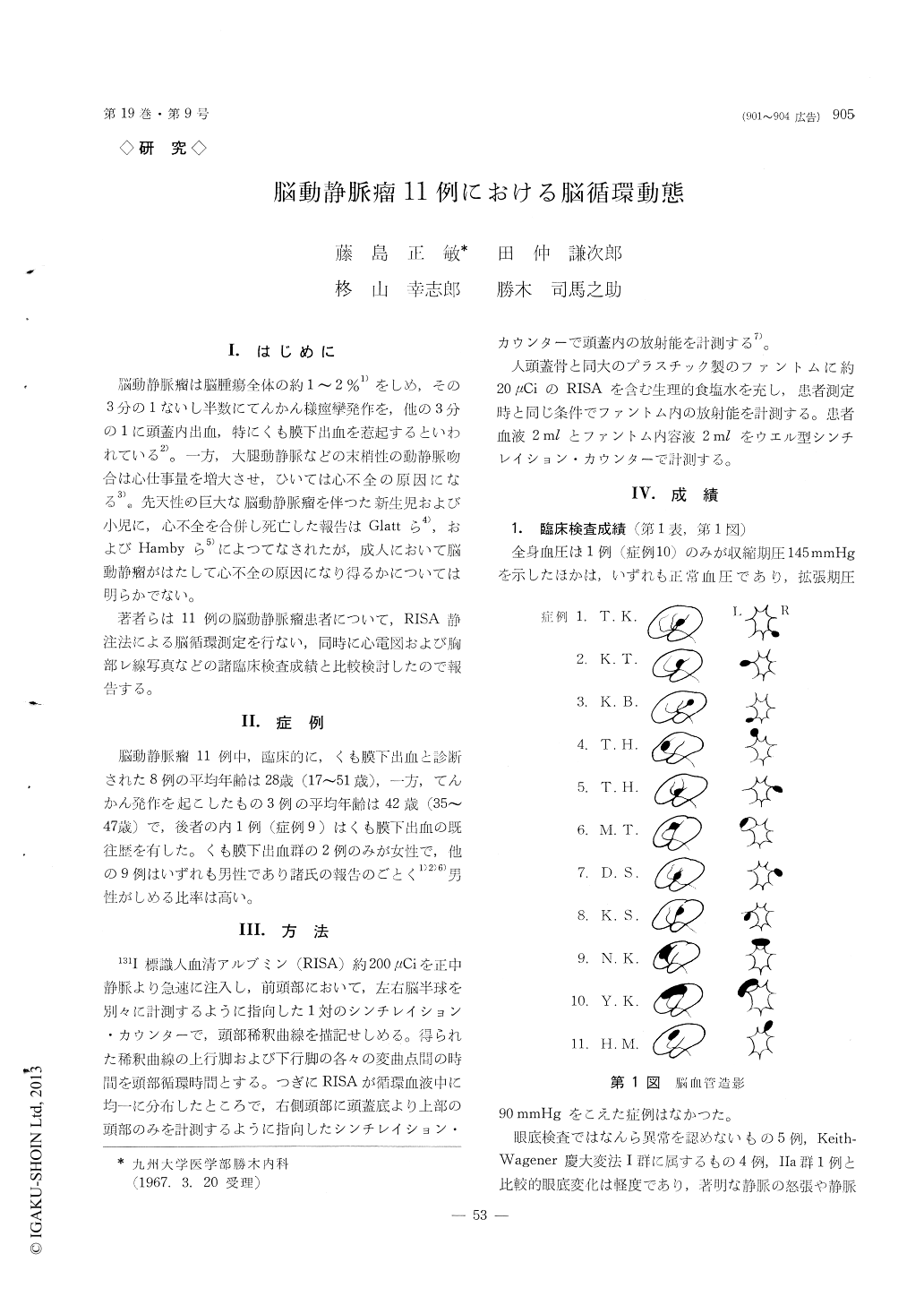
著者らは11例の脳動静脈瘤患者について,RISA静注法による脳循環測定を行ない,同時に心電図および胸部レ線写真などの諸臨床検査成績と比較検討したので報告する。
Measurements of transit time (TT), cranial blood volume (CBV) and cranial blood flow (CBF) were made using the method by external counting of in-travenously injected RISA in 11 cases with cerebral arteriovenous malformation.
TT was shortened in 3 cases, CBV and CBF were significantly increased in 4 and 5 cases respectively, comparied with those in 34 normotensive control subjects.
In 3 epileptic subjects with large cerebral A-V malformation locatad in the frontoparietal region, CBF was markedly increased. However, each para-meter of brain circulation was varied in 8 cases mani-fested subarachnoid hemorrhage. Cerebral hemody-namic alterations were considered to depend on the size of the A-V malformation and its location in the arterial tree.
Though increase in CBF was marked in 4 cases with the left ventricular hypertrophy shown by ele-ctrocardiogram and/or with the cardiac enlarge-ment revealed by chest X-ray film, conjestive heart failure was observed in none of 11 cases. The in-creased CBF and cardiac output were considered the major factors contributing to the cardiac change.

Copyright © 1967, Igaku-Shoin Ltd. All rights reserved.


