Japanese
English
- 有料閲覧
- Abstract 文献概要
- 1ページ目 Look Inside
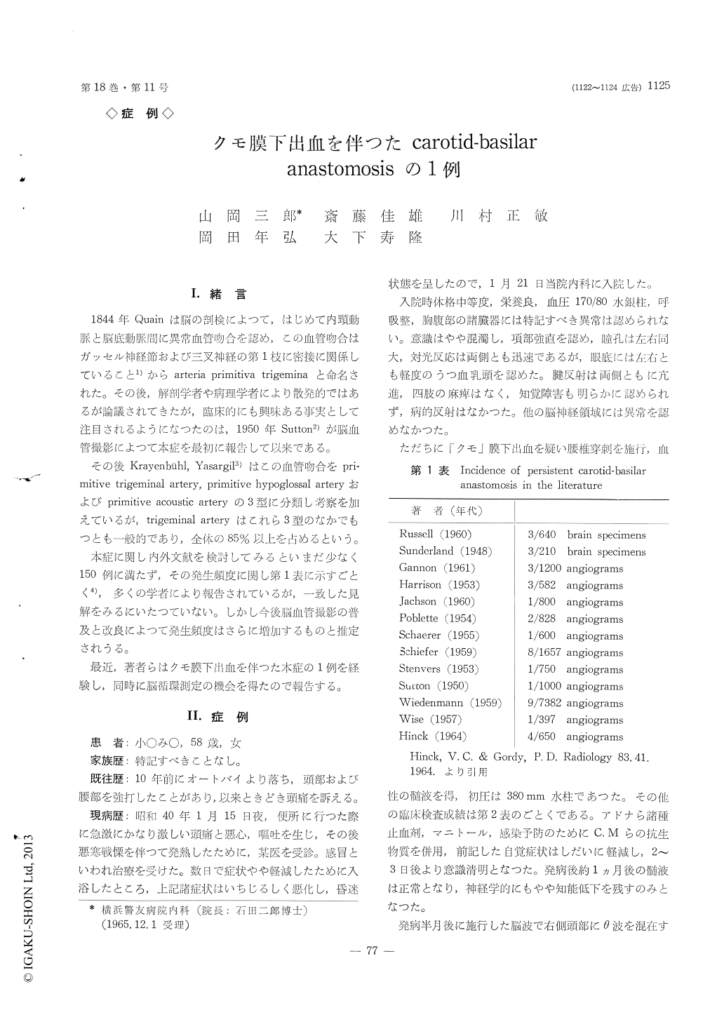
I.緒言
1844年Quainは脳の剖検によつて,はじめて内頸動脈と脳底動脈間に異常血管吻合を認め,この血管吻合はガッセル神経節および三叉神経の第1枝に密接に関係しているにと1)からarteria primitiva trigeminaと命名された。その後,解剖学者や病理学者により散発的ではあるが論議されてきたが,臨床的にも興味ある事実として注目されるようになつたのは,1950年Sutton2)が脳血管撮影によつて本症を最初に報告して以来である。
その後Krayenbuhl, Yasargil3)はこの血管吻合をpri—mitive trigeminal artery, primitive hypoglossal arteryおよびprimitive acoustic arteryの3型に分類し考察を加えているが,trigeminal arteryはこれら3型のなかでもつとも一般的であり,全体の85%以上を占めるという。
A case of 58 years old housewife with the diag-nosis of trigeminal artery is reported.
This patient was admitted to the Keiyu Hospital with a complaint of severe occipital headache and confusional state. One week prior to admission, she suddenly started to have severe headache, nausea, vomiting and fever, and visited a local doctor who reated, her as a cold. Since her symptoms had im-proved by his treatment, 3days (prior to admission) p. t. a. she took a bath, and subsequently these sym-ptoms are worsened and she fell into the confusional status.
Physical examination on admission.
Patient is well nourished, moderately developed. Blood pressure is 170-80. Respiration is regular. No abnormality in chest and abdomen is found. Her consciousness is slightly disturbed, and nuc-hal rigidity is present. Optic fundi showed bilateral early papilloedema, deep tendon reflexes are hyper-active throughout, without pathologic reflex. There is no definite weakness or sensory impairment. Cra-nial nerves revealed no other abnormality. Lumbar puncture was performed, immediately after admis-sion, showed bloody spinal fluid with pressure of 380 mm H2O.
Right carotid-arteriography, which was performed one month after the onset of this accident, demons-trated abnormal vessel communicating between the basilar artery and the carotid siphon. This is the carotidbasilar anastomosis which is called persistent primitive trigeminal artery. There is no aneurysma, displacement of the vessel or arteriovenous fistula in the cerebral hemisphere. However, it is suggested that there might be abnormal vasculature around the right posterior cerebral artery.
Right subclavian arteriography also visualized pri-mitive trigeminal artery which may be supplied by the vertebral artery.
Cerebral circulation and hemodynamics were me-asured by N2O method and the following results were obtained.
CBF is 53. 7 which is within normal limits,
CVR is 2.62 which is increased, and
CMRO2 2. 03, decreased.
Since there is no definite evidence of hemorrhagic origin, the source of bleeding is still questionable to point out in this case. However, according to Saltzman and his associates it is assumed to be origin of hemo-rrhage that this anastomosis sometimes revealed wi-despread media defect in histologic study. This theory could be applied for this case.
Cerebral circulation were in the range of normal values, on the contrary to arteriovenous fistula which should be increased. As regard to the decreased CMRO2, it could be explained that this patient had still shown slight dementia at the time of measure-ment.

Copyright © 1966, Igaku-Shoin Ltd. All rights reserved.


