Japanese
English
- 有料閲覧
- Abstract 文献概要
- 1ページ目 Look Inside
I.緒言
脳血管撮影法は1927年E.Monizにより創められたが,その後種々改良を加えられ,今日では連続撮影装置の併用と共に,その応用範囲は一段と拡大されその診断価値はますます高められつつある状態である1)2)5)。
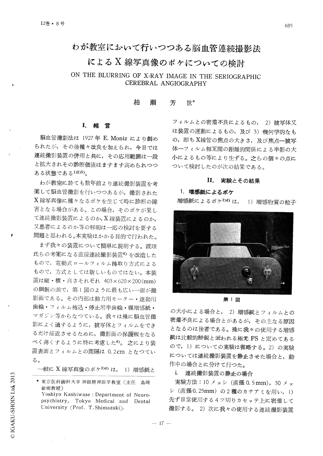
わが教室に於ても数年前より連続撮影装置を考案して脳血管撮影を行いつつあるが,撮影されたX線写真像に種々なるボケを生じて時に診断の障害となる場合がある。この場合,そのボケが果して連続撮影装置によるのか,X線装置によるのか,又患者によるのか等の解明は一応の検討を要する問題と思われる。本実験はかかる目的で行われた。
This study concerns itself with the blur-ring of the serial angiogram which was oc-casionally encountered and hurt the diagnos-tic value.
The result are as follows:
1) The blurring is caused by incompletene- ss of contact between intensifying screens and films. While the film is moving, the incompleteness of contact becomes more larger.
2) The vibration of the apparatus while working enhances the blurring of the ro- eutgenographic image.
3) An object of 0.5mm in diameter is enou- gh to produce the image on the roentgeno- gram.
4) The cause of the blurring of an object over 0.5 mm in diameter should be ascri- bed to the motion of the patient, technical failure of angiographer, and unsatisfactory contrast media.

Copyright © 1960, Igaku-Shoin Ltd. All rights reserved.


