Japanese
English
- 有料閲覧
- Abstract 文献概要
- 1ページ目 Look Inside
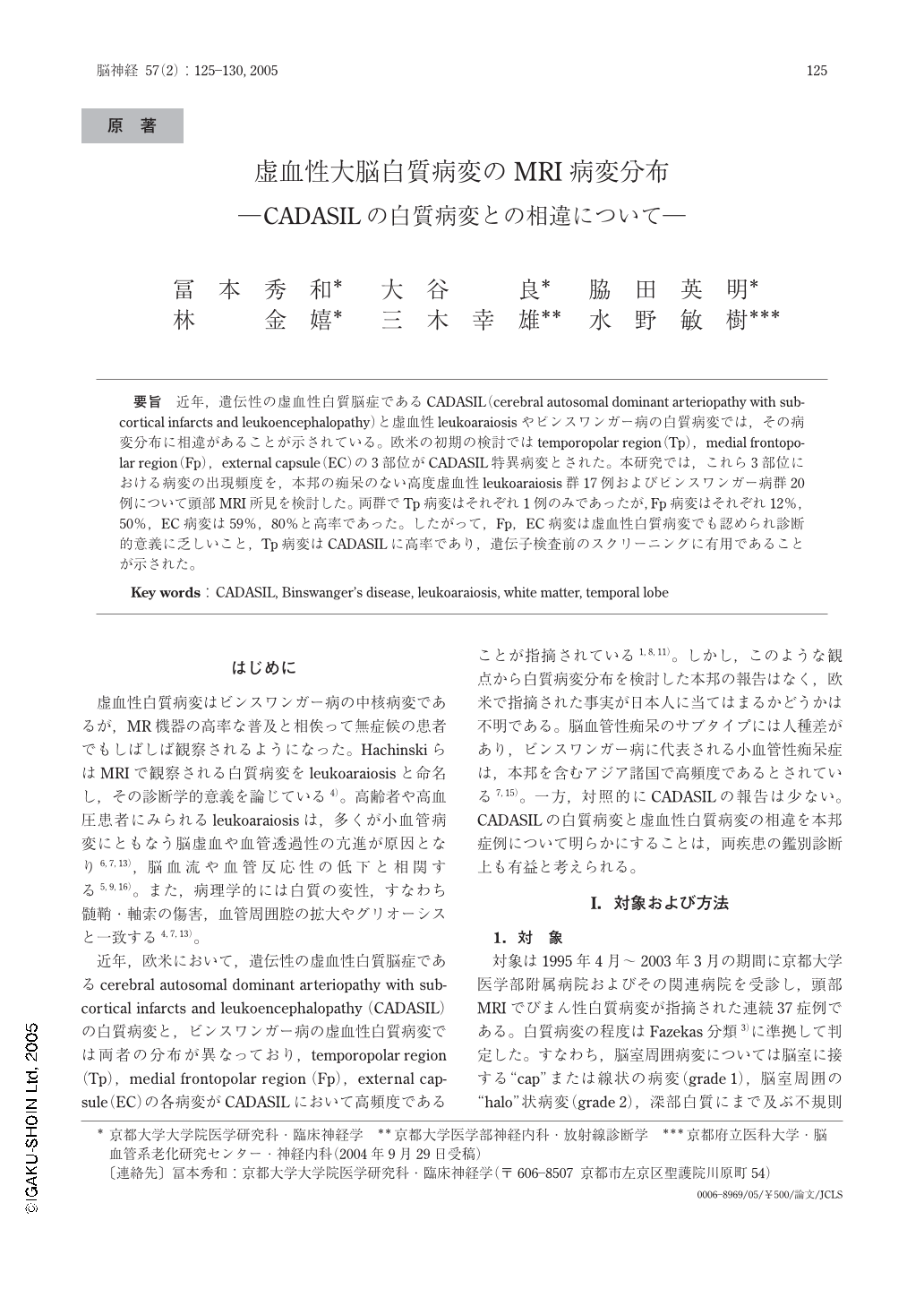
要旨 近年,遺伝性の虚血性白質脳症であるCADASIL(cerebral autosomal dominant arteriopathy with subcortical infarcts and leukoencephalopathy)と虚血性leukoaraiosisやビンスワンガー病の白質病変では,その病変分布に相違があることが示されている。欧米の初期の検討ではtemporopolar region(Tp),medial frontopolar region(Fp),external capsule(EC)の3部位がCADASIL特異病変とされた。本研究では,これら3部位における病変の出現頻度を,本邦の痴呆のない高度虚血性leukoaraiosis群17例およびビンスワンガー病群20例について頭部MRI所見を検討した。両群でTp病変はそれぞれ1例のみであったが,Fp病変はそれぞれ12%,50%,EC病変は59%,80%と高率であった。したがって,Fp,EC病変は虚血性白質病変でも認められ診断的意義に乏しいこと,Tp病変はCADASILに高率であり,遺伝子検査前のスクリーニングに有用であることが示された。
Previously, the distribution of white matter lesions in CADASIL has been reported to be distinct from those in patients with ischemic leukoaraiosis and Binswanger's disease. In earlier European studies, diagnostic significance of white matter lesions in the temporopolar region(Tp), medial frontopolar region(Fp) and external capsule(EC) was stressed in diagnosing CADASIL. More recently, however, high sensitivity and specificity of Tp lesions have been demonstrated. In Japan, prevalence of CADASIL is lower, and those of ischemic leukoaraiosis and Binswanger's disease, likely related to small artery disease, are much higher than in Caucasian countries. Therefore, we examined the frequencies of CADASIL-associated lesions in 17 non-demented patients with ischemic leukoaraiosis and 20 patients with Binswanger's disease. The Binswanger's disease group showed a significantly lower scores for Hasegawa Dementia Rating Scale Revised(HDSR) and a higher prevalence of hypertension, compared to the ischemic leukoaraiosis group. There was only 1 patient with Tp lesions in each group, while Fp lesions were found in 12% and 50% in the ischemic leukoaraiosis group and Binswanger's disease group, respectively, and EC lesions in 59% and 80%. These results indicated that Tp lesions were useful diagnostic marker in diagnosing CADASIL, whereas Fp and EC lesions were non-specifically observed.
(Received : September 29, 2004)

Copyright © 2005, Igaku-Shoin Ltd. All rights reserved.


