Japanese
English
- 有料閲覧
- Abstract 文献概要
- 1ページ目 Look Inside
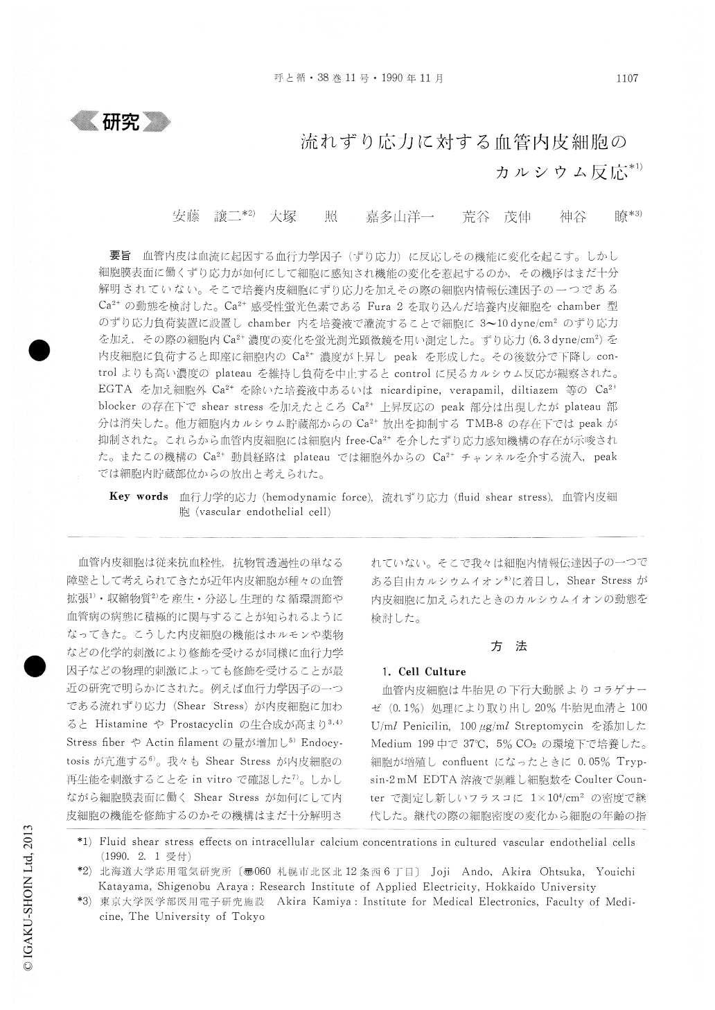
血管内皮は血流に起因する血行力学因子(ずり応力)に反応しその機能に変化を起こす。しかし細胞膜表面に働くずり応力が如何にして細胞に感知され機能の変化を惹起するのか,その機序はまだ十分解明されていない。そこで培養内皮細胞にずり応力を加えその際の細胞内情報伝達因子の一つであるCa2+の動態を検討した。Ca2+感受性蛍光色素であるFura 2を取り込んだ培養内皮細胞をchamber型のずり応力負荷装置に設置しchamber内を培養液で灌流することで細胞に3〜10dyne/cm2のずり応力を加え,その際の細胞内Ca2+濃度の変化を蛍光測光顕微鏡を用い測定した。ずり応力(6.3 dyne/cm2)を内皮細胞に負荷すると即座に細胞内のCa2+濃度が上昇しpeakを形成した。その後数分で下降しcon-trolよりも高い濃度のplateauを維持し負荷を中止するとcontrolに戻るカルシウム反応が観察された。EGTAを加え細胞外Ca2+を除いた培養液中あるいはnicardipine,verapamil,diltiazem等のCa2+blockerの存在下でshear stressを加えたところCa2+上昇反応のpeak部分は出現したがplateau部分は消失した。他方細胞内カルシウム貯蔵部からのCa2+放出を抑制するTMB−8の存在下ではpeakが抑制された。これらから血管内皮細胞には細胞内free-Ca2+を介したずり応力感知機構の存在が示唆された。またこの機構のCa2+動員経路はplateauでは細胞外からのCa2+チャンネルを介する流入,peakでは細胞内貯蔵部位からの放出と考えられた。
Vascular endothelial cells are known to modulate their functions in response not only to humoral stimuli but also to such physical stimuli as fluid shear stress generated by blood flow. However, the mechanisms by which the hemodynamic force acts on endothelial cells are not yet well understood. We have studied how endothelial cells recognizethe shear stress and mediate it to intracellular or-ganelles. Cultured monolayers of bovine aortic en-dothelial cells loaded with the highly fluorescent Ca++-sensitive dye Fura 2 were exposed to different levels of fluid shear stress in a specially designed flow chamber and simultaneous changes in intracel-lular-free Ca++ concentration were measured using photometric fluorescence microscopy.
Application of shear stress to cells by fluid perfu-sion led to an immediate several-fold increase in Ca++ concentration within 1 min, followed by a rapid decline, and finally a plateau somewhat higher than control levels during the entire period of the stress application. The early part of the response, but no plateau, was observed even in Ca++-free me-dium added with 2mM EDTA, and in the presence of calcium antagonists (e. g. 2x10⁻⁵M nicardipine), Thus, endothelial cells may have a flow-sensing property which recognize the shear stress on the membrane as a stimulus and mediates the signal to increase intracellular free Ca++ which is a major component of the internal signalling system of the cell.

Copyright © 1990, Igaku-Shoin Ltd. All rights reserved.


