Japanese
English
- 有料閲覧
- Abstract 文献概要
- 1ページ目 Look Inside
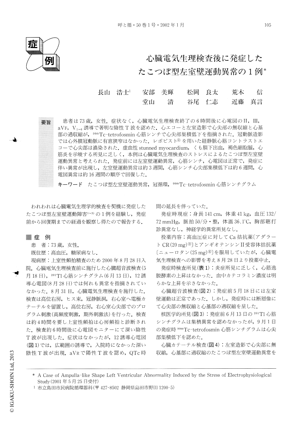
要旨 患者は73歳、女性.症状なく、心臓電気生理検査終了の6時間後に心電図のII・III、aVF,V1〜6誘導で著明な陰性T波を認めた.心エコーと左室造影で心尖部の無収縮と心基部の過収縮が,99mTc-tetrofosmin心筋シンチで心尖部集積低下を指摘された.冠動では心外膜冠動脈に有意狭窄はなかった.レボビスト®を用いた経静脈心筋コントラストエコーで心尖部は濃染された.虚血性stunned myocardium,くも膜下出血,褐色細胞腫,心筋炎を示唆する所見に乏しく,本例は心臓電気生理検査のストレスによるたこつぼ型左室壁運動異常と考えられた.発症前には左室壁運動異常,心筋シンチ,心電図は正常で,発症に伴い異常が出現し,左室壁運動異常は約3週間,心筋シンチ心尖部集積低下は約6週間,心電図異常は約16週間の順序で回復した.
We present a case of a 73-year-old woman showing the development of an ampulla-like shape left ventricular wall motion abnormality associated with a diagnostic electrophysiological study, as outlined elsewhere, to evaluate supuraventricular tachycardia. Before the electrophysiological study, electrocardiogram, left ventricular wall motion and thallium-201 myocardial imaging were confirmed as normal. Although she had no symptoms, electrocardiogram obtained 6 hours after the electrophysiological study showed deep inverted T waves with prolonged QT intervals in the II, III, aVF and V1-6leads. A balloon-like akinetic area at the apex and hypercontraction of the basal segment of the left ventricle was observed by echocardiography and left ventriculogram. Technetium-99m tetrofosmin myocardial imaging revealed decreased uptake at the apex of the left ventricle. Coronary angiogram demonstrated no significant organic stenosis in the epicardial coronary arteries. Echo enhancement using Levovist was observed at the akinetic area of the ventricle. There was no evidence of ischemic stunned myocardium, subarachnoid hemorrhage, pheochromocytoma crisis or myocarditis. Thus, the cause of the left ventricular wall motion abnormality was considered to be the stress of the electrophysiological study.
The ampulla-like shape of the left ventricular wall motion, decreased uptake of myocardial technetium-99m tetrofosmin at the apex and the abnormalities on electrocardiogram returned to normal at 3, 6 and 16 weeks, respectively. Because there was no evidence of abnormality in her coronary circulation, the cause of transient scintigraphic abnormality was considered to be mitochondrial injury.
We reported the clinical course in a case showing an ampulla-like shape left ventricular wall motion abnormality. Further investigation is needed to understand the mechanism of this entity.

Copyright © 2002, Igaku-Shoin Ltd. All rights reserved.


