Japanese
English
- 有料閲覧
- Abstract 文献概要
- 1ページ目 Look Inside
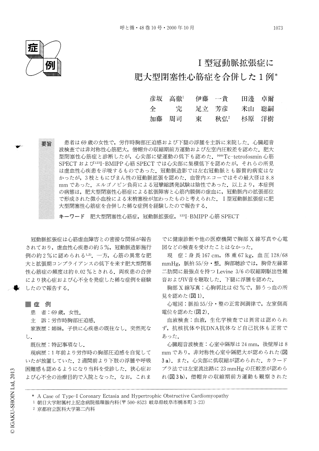
患者は69歳の女性で,労作時胸部圧迫感および下腿の浮腫を主訴に来院した.心臓超音波検査では非対称性心筋肥大,僧帽弁の収縮期前方運動および左室内圧較差を認めた.肥大型閉塞性心筋症と診断したが,心尖部に壁運動の低下も認めた.99mTC-tetrofosmin心筋SPECTおよび123I-BMIPP心筋SPECTでは心尖部に集積低下を認めたが,それらの所見は虚血性心疾患を示唆するものであった.冠動脈造影では左右冠動脈とも器質的病変はなかったが,3枝ともにびまん性の冠動脈拡張を認めた.血管内エコーではその最大径は8.8mmであった.エルゴノビン負荷による冠攣縮誘発試験は陰性であった.以上より,本症例の病態は,肥大型閉塞性心筋症による拡張障害と心筋内膜側の虚血に,冠動脈内の拡張部位で形成された微小血栓による末梢塞栓が加わったものと考えられた.I型冠動脈拡張症に肥大型閉塞性心筋症を合併した稀な症例を経験したので報告する.
A 69 year-old-woman developed chest oppression oneffort and leg edema. Echocardiography showed asym-metric septal hypertrophy, systolic anterior motion ofthe mitral valve leaflet, left ventricular pressure gradi-ent and hypokinesis at the apex. These echocardiogra-phic findings, except for hypokinesis at the apex, werecompatible with hypertrophic obstructive car-diomyopathy. 99mTc-tetrofosmin myocardial SPECTshowed reduced uptake in the apex, and 123I-BMIPPmyocardial SPECT showed reduced uptake at the anter-oseptal wall and apex. These SPECT findings werecompatible with ischemic heart disease rather than withhypertrophic obstructive cardiomyopathy. Coronaryangiography showed no organic stenosis, but diffusecoronary ectasia was noted in three vessels. Intravas-cular ultrasound revealed remarkable coronary ectasia,and the maximal diameter was 8.8mm. These findingssuggest that the etiology in this patient might be ex-plained by hypertrophic obstructive cardiomyopathyand microembolization due to coronary ectasia.

Copyright © 2000, Igaku-Shoin Ltd. All rights reserved.


