Japanese
English
- 有料閲覧
- Abstract 文献概要
- 1ページ目 Look Inside
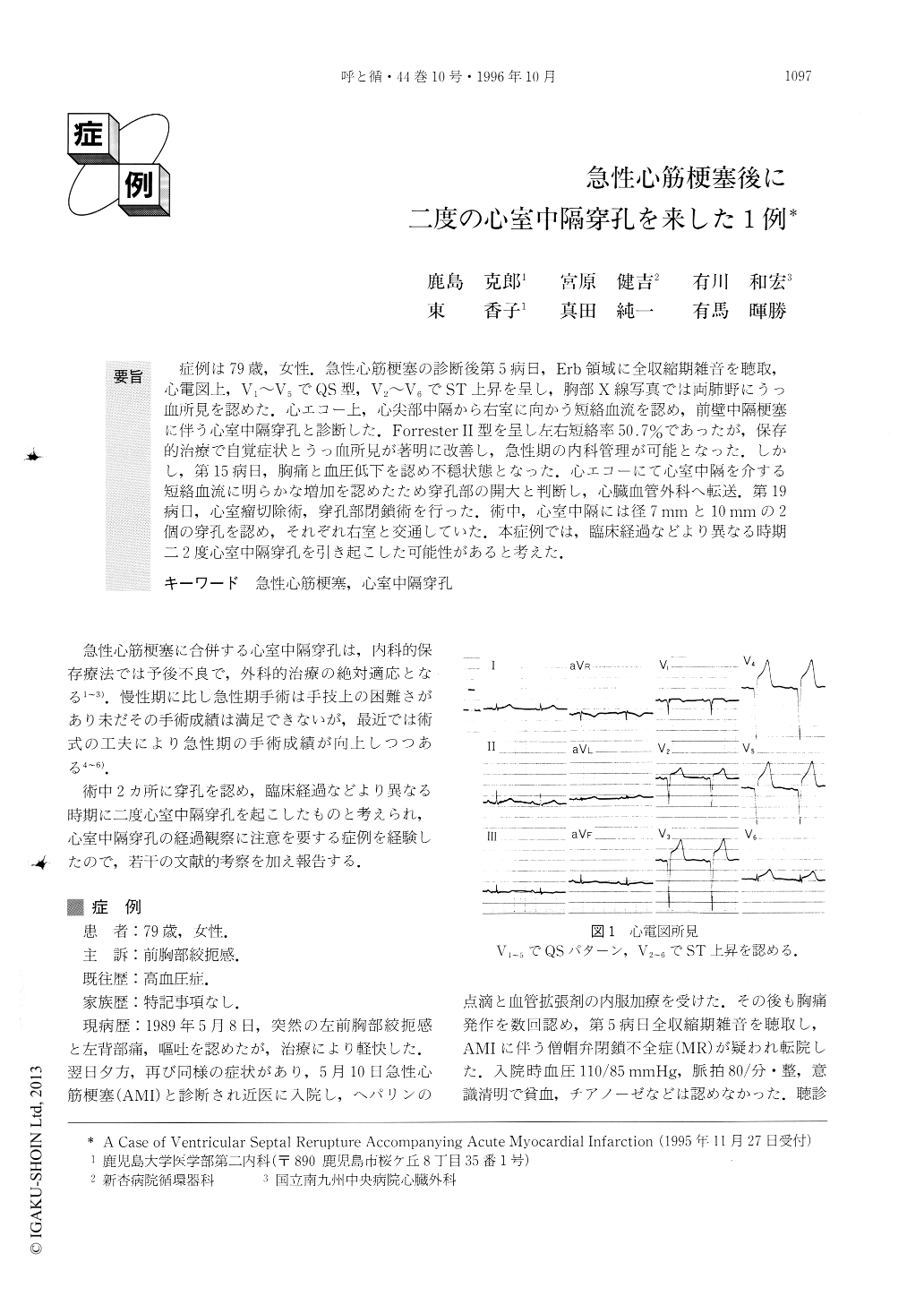
症例は79歳,女性.急性心筋梗塞の診断後第5病日,Erb領域に全収縮期雑音を聴取,心電図上,V1〜V5でQS型,V2〜V6でST上昇を呈し,胸部X線写真では両肺野にうっ血所見を認めた.心エコー上,心尖部中隔から右室に向かう短絡血流を認め,前壁中隔梗塞に伴う心室中隔穿孔と診断した.Forrester II型を呈し左右短絡率50.7%であったが,保存的治療で自覚症状とうっ血所見が著明に改善し,急性期の内科管理が可能となった.しかし,第15病日,胸痛と血圧低下を認め不穏状態となった.心エコーにて心室中隔を介する短絡血流に明らかな増加を認めたため穿孔部の開大と判断し,心臓血管外科へ転送。第19病日,心室瘤切除術,穿孔部閉鎖術を行った.術中,心室中隔には径7mmと10mmの2個の穿孔を認め,それぞれ右室と交通していた.本症例では,臨床経過などより異なる時期二2度心室中隔穿孔を引き起こした可能性があると考えた.
The patient was a 79-year-old woman. She was admitted because of acute myocardial infarction. On the 5th hospital day, systolic murmur was heard. An electrocardiogram showed QS type in V1-V5 leads and ST elevation in V2-V6 leads, and pulmonary conges-tions were detected in both lung fields on chest x-ray films. Short-circuit blood flow running from the apical region of the septum to the right ventricle was observed by the color Doppler method. We made a diagnosis of ventricular septal perforation accompanying anterose-ptal infarction in the patient. Forrester II type was seen and the left-to-right shunt rate was 50.7 %; conserva-tive treatment markedly improved subjective symptoms and pulmonary congestions, and internal-medical con-trol of the disease became possible. However, chest pain developed on the 15th hospital day,the patient's blood pressure decreased at the same time and restless-ness occurred. Although there was no change on the electrocardiogram, a clear increase in the amount of blood shunted through the interventricular septum was observed by the color Doppler method, so that we jud-ged the event to be enlargement of the perforation, and transferred the patient to the Department of Cardiovas-cular Surgery. Resection of the ventricular aneurysm and closure of perforation was performed on the 19th hospital day. During the operation, 2 perforations, 7mm and 10mm in diameter, respectively, were observed in the ventricular septum and they communicated with the right ventricle individually. On the basis of our observa-tions of the clinical course of this case, we considered that ventricular septal perforation might have occurred twice at different times in this patient.

Copyright © 1996, Igaku-Shoin Ltd. All rights reserved.


