Japanese
English
- 有料閲覧
- Abstract 文献概要
- 1ページ目 Look Inside
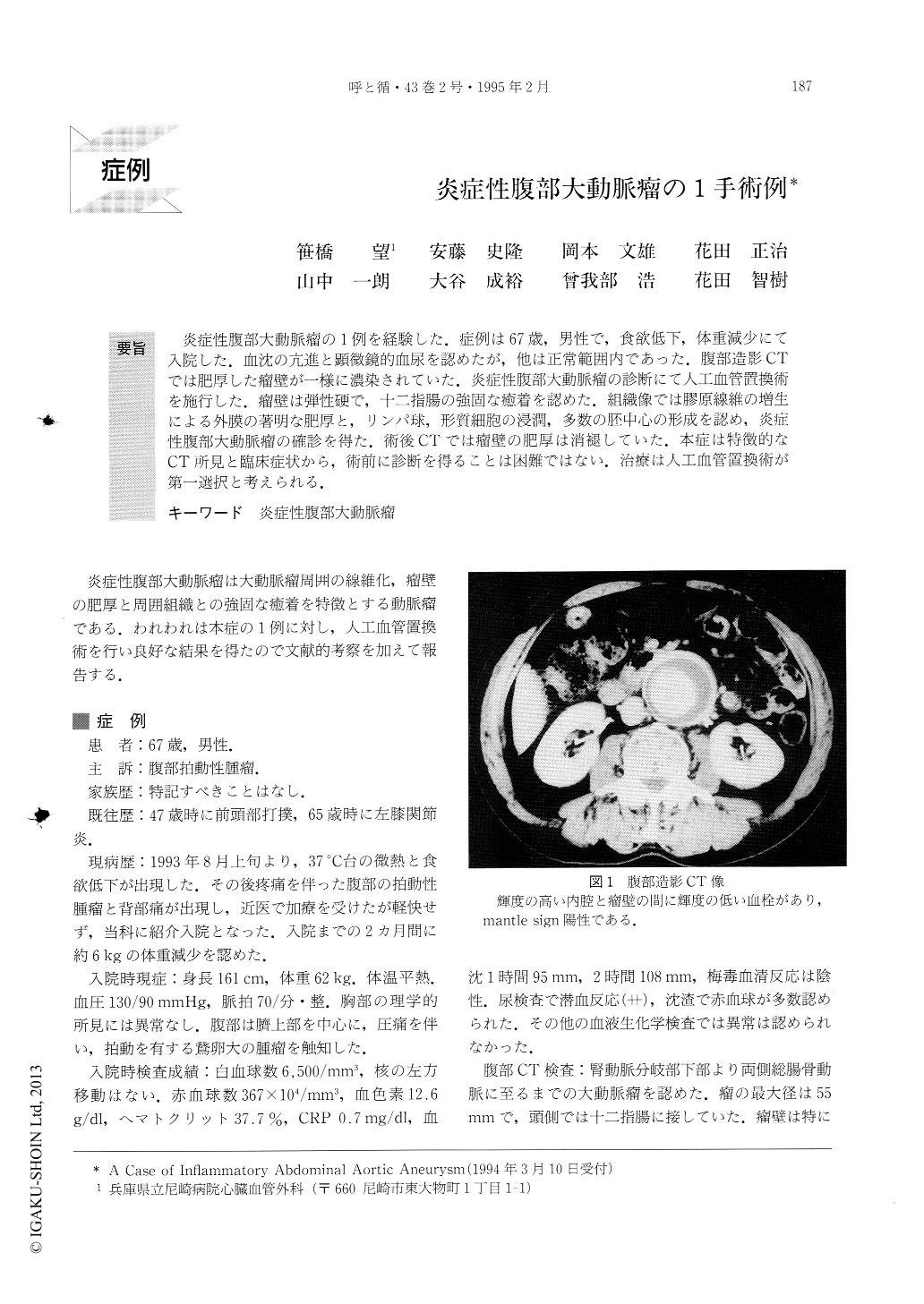
炎症性腹部大動脈瘤の1例を経験した.症例は67歳,男性で,食欲低下,体重減少にて入院した.血沈の亢進と顕微鏡的血尿を認めたが,他は正常範囲内であった.腹部造影CTでは肥厚した瘤壁が一様に濃染されていた.炎症性腹部大動脈瘤の診断にて人工血管置換術を施行した.瘤壁は弾性硬で,十二指腸の強固な癒着を認めた.組織像では膠原線維の増生による外膜の著明な肥厚と,リンパ球,形質細胞の浸潤,多数の胚中心の形成を認め,炎症性腹部大動脈瘤の確診を得た.術後CTでは瘤壁の肥厚は消褪していた.本症は特徴的なCT所見と臨床症状から,術前に診断を得ることは困難ではない.治療は人工血管置換術が第一選択と考えられる.
A case of 67-year-old man with inflammatory abdom-inal aortic aneurysm was reported. Two months before admission he complained of anorexia, pulsatile abdomi-nal mass, and body weight loss.
Erythrocyte sedimentation rate was 95mm/hr, and urinalysis revealed microhematuria. A CT scan demon-strated a thickened aneurysm wall which enhanced after contrast medium injection. After the diagnosis of the inflammatory abdominal aortic aneurysm was made, an operation was performed. At surgery, the aneurysm wall was found to be thickening and its elasticity had hardened. The densely adherent duode-num was not completely mobilized. A replacement of the aneurysm wall was accomplished with a bifurcated graft in the standard manner. The patient was dischar-ged without complications one month after the opera-tion. Histopathological findings showed marked col-lagen fibrosis in the adventitia, infiltration of lymphocytes and plasma cells, and germinal centers. Three months after the operation, the CT scan revealed significant regression of the wall thickening which was enhanced less than preoperatively. It is possible to make a diagnosis of an inflammatory abdominal aortic aneurysm preoperatively in the presence of typical CT findings and symptoms. Once the diagnosis is made, a graft replacement should be preferred.

Copyright © 1995, Igaku-Shoin Ltd. All rights reserved.


