Japanese
English
- 有料閲覧
- Abstract 文献概要
- 1ページ目 Look Inside
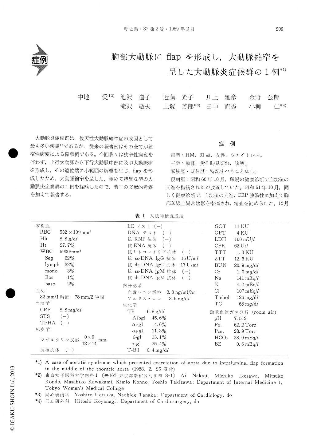
大動脈炎症候群は,後天性大動脈縮窄症の成因として最も多い疾患1)であるが,従来の報告例はその全てが狭窄性病変による縮窄例である。今回我々は狭窄性病変を伴わず,上行大動脈から下行大動脈中部に及ぶ大動脈瘤を形成し,その遠位端に小範囲の解離を生じ,flapを形成したため,大動脈縮窄を呈した,極めて特異な型の大動脈炎症候群の1例を経験したので,若干の文献的考察を加えて報告する。
We present a rare case of aortitis syndrome asso-ciated with dilatation of aorta and coarctation-like effect due to the intraluminal flap formation originated from dissected wall of the aorta.
A 31-year-old woman was admitted to our hospitalcomplaining of shortness of breath, palpitation and cough. On admission, her physical status showed con-gestive heart failure and hypertension of upper extre-mities and hypotension of lower extremities. Bruits were audible over the neck, the anterior chest and the back. Serological studies showed active inflamma-tion. Chest X-ray film showed upper mediastinal widening, cardiomegaly and pulmonary edema. Aortitis syndrome was strongly suggested by these clinical findings, so that prednisolone therapy was started on 3rd hospital day. Special examinations were performed several days later when inflammato-ry changes showed a tendency to improve.
Chest CT scan, RI angiography and MRI studies showed an aneurysmal dilatation from the ascending aorta to the mid-thoracic aorta. Aortography demo-nstrated a flap at the terminal portion of this aneury-smal dilatation and grade II (Sellars) aortic regurgi-tation. There was a pressure difference of 80 mmHg between the parts abutting cranial and caudal sides of the flap.
A surgical operation was, then, performed to cor-rect the pressure difference. The disscted wall was extruded toward the aortic lumen creating a flap (2 cm in length). This flap was resected and an arti-ficial graft was inserted. Histologically, the flap con-sisted of adventitia, media and intima. The adven-titia and media were accompaniied by infiltration of inflammatory cells and the elastic fibers of the media were torn diffusely showing that the dissection was due to mesoaortitis.
Catheterization and aortography were performed after the operation which confirmed that it had been made successfully.

Copyright © 1989, Igaku-Shoin Ltd. All rights reserved.


