Japanese
English
- 有料閲覧
- Abstract 文献概要
- 1ページ目 Look Inside
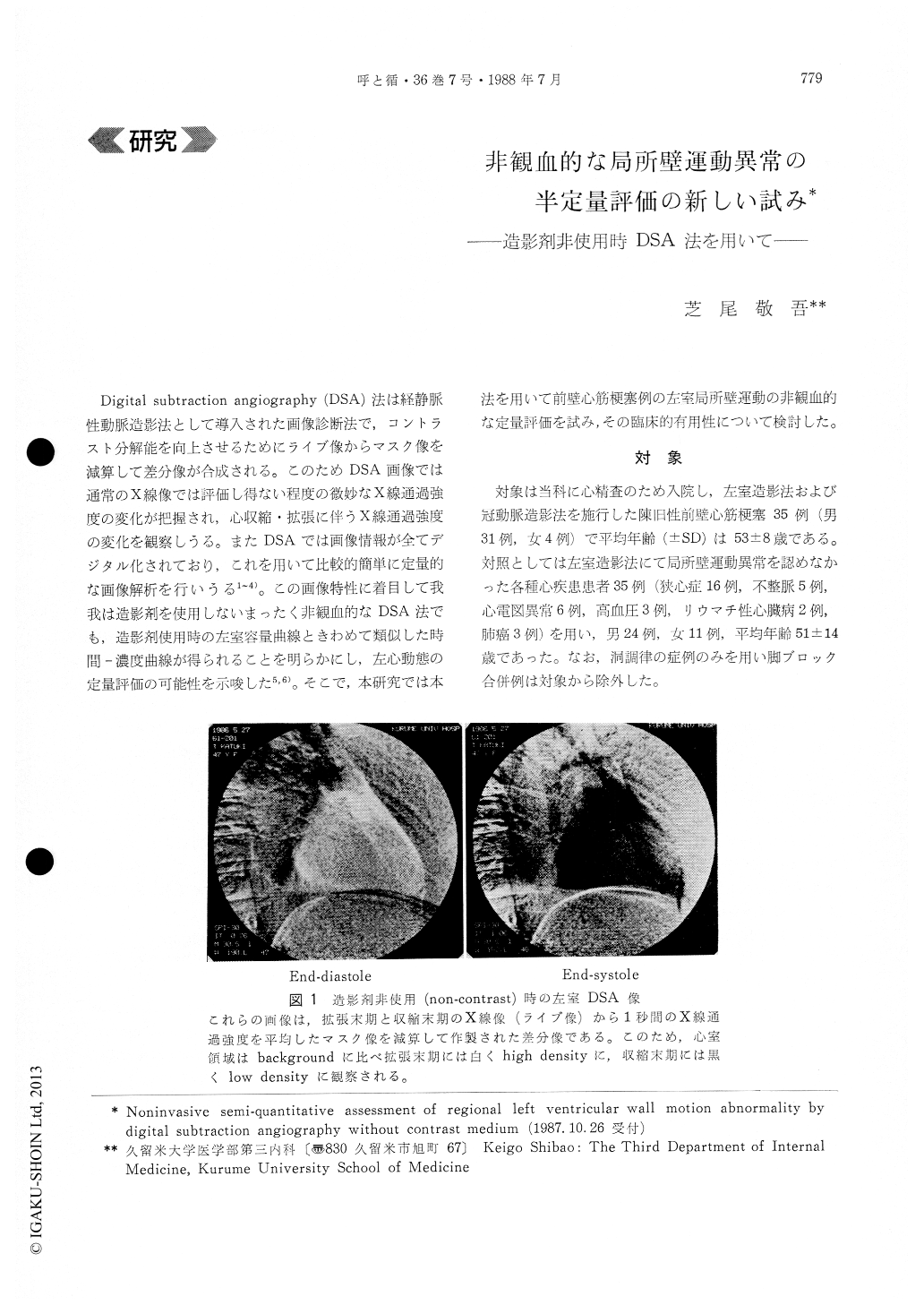
Digital subtraction angiography (DSA)法は経静脈性動脈造影法として導入された画像診断法で,コントラスト分解能を向上させるためにライブ像からマスク像を減算して差分像が合成される。このためDSA画像では通常のX線像では評価し得ない程度の微妙なX線通過強度の変化が把握され,心収縮・拡張に伴うX線通過強度の変化を観察しうる。またDSAでは画像情報が全てデジタル化されており,これを用いて比較的簡単に定量的な画像解析を行いうる1〜4)。この画像特性に着目して我我は造影剤を使用しないまったく非観血的なDSA法でも,造影剤使用時の左室容量曲線ときわめて類似した時間—濃度曲線が得られることを明らかにし,左心動態の定量評価の可能性を示唆した5,6)。そこで,本研究では本法を用いて前壁心筋梗塞例の左室局所壁運動の非観血的な定量評価を試み,その臨床的有用性について検討した。
Since digital subtraction angiography (DSA) has been developed to enhance the contrast to back-ground signal, the method appeared to be capable of assessing a small change in X-ray absorption during a single cardiac cycle without contrast medium (CM). With this advantage, we have deve-loped a new noninvasive method to evaluate regional left ventricular (LV) function. DSA images of the LV without and with CM (non-contrast and contrast DSA), were obtained from 35 patients with anterior myocardial infarction (AMI) and from 35 control subjects. Using an image-processing computer, re-gional LV time-density curves were constructed for one cardiac cycle. Regional LV time-density curves from non-contrast DSA presented a similar pattern to those from contrast DSA. The amplitude of re-gional LV time-density curves in patients with AMI decreased along with increasing severity of asynergy assessed by conventional left ventriculography (LVG). In an attempt of semi-quantitative evaluation with non-contrast DSA, regional wall motion index (RWI) in the 6 segments of the LV was calculated from segmental density change devided by maximal seg-mental density change. RWI showed a good cor-relation with regional ejection fraction (REF) ob-tained from intravenous contrast DSA. As compared with control subjects, patients with AMI have sig-nificantly lower RWIs in the antero-lateral and apical regions. RWI decreased with increasing severity of regional wall motion abnormality in the conventional LVG. The diagnostic accuracy of RWI was comparable to that of REF derived from con-trast DSA.
Thus, the present study indicated that the com-puter analysis of non-contrast DSA image is avaluable noninvasive method for semi-quantitative assessment of regional LV function.

Copyright © 1988, Igaku-Shoin Ltd. All rights reserved.


