Japanese
English
- 有料閲覧
- Abstract 文献概要
- 1ページ目 Look Inside
放射線照射による心障害には,心嚢液貯留を伴う急性心外膜炎やその慢性化があるが,なかには,収縮性心外膜炎に移行する例,心内膜と心筋に線維化を伴う重篤な例も報告されている1〜4)。慢性収縮性心外膜炎と,心内膜ないし心筋の線維化による拘束型心筋症との鑑別は,しばしば困難である。放射線心障害では,この2つの病態が共存することがあるので,収縮性心外膜炎に心内膜ないし心筋の線維化を合併し得る症例では,あらかじめ心筋生検を行ない,心内膜や心筋の病変を検索し,その後に心膜剥離術の適否を考慮十ることが大明である。
今回我々は,肋骨腫瘍摘出後の放射線照射によって,急性心外膜炎を呈し,その後に心嚢液貯留を伴う慢性収縮性心外膜炎に陥り,心筋生検により,軽度の心筋病変を認めた症例を経験したので報告し,あわせて放射線心障害についての文献的考察を行なった。
A 25-year-old female was admitted with shortness of breath and abdominal swelling. Six years before the admission, she had received resection of 7th to 9th left ribs and subsequent radiation of 5,000 rads to the thorax for the treatment of rib osteo-blastoma. One year after the radiation, marked pericardial effusion associated with acute pericarditis was observed and was improved by digitalization and diuretic therapy. Since two years after the radiation, she had felt easy fatigability, swelling of face and foot, and transient faintness on more than 10 meters running.
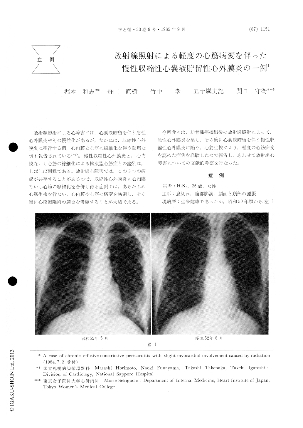
On admission, chest X-ray photograph showed increased pulmonary vascularity without cardiac enlargement. Electrocardiogram indicated systolic right ventricular strain, mitral P, and nonspecifiic S-T segment depression in left precordial leads. Two-dimensional echocardiography presented peri-cardial effusion with posterior pericardial thicken-ing, while M-mode echocardiography showed diasto-lic posterior movement of interventricular septum and diastolic flattening of left ventricular posterior wall.
Cardiac catheterization revealed marked elevation of mean right atrial pressure, pulmonary arterial diastolic pressure, right and left ventricular end-diastolic pressure, accompanied with their equaliza-don. In addition, pressure waves of right and left ventricle showed diastolic dip and plateau. Phono-cardiogram and apexcardiogram presented peri-cardial knock sound and systolic retraction, respec-tively. Cardiac angiography showed diastolic restric-tion of left ventricle without any stenosis of coro-nary artery. Computed tomography (CT) of the chest revealed thickening of anterior and left lateral pericardium with expansion of inferior vena cava, and abdominal CT revealed ascites with slight enlargements of liver and spleen.
From above obtained data, chronic effusive-con-strictive pericarditis, which was attributed to radia-tion, was strongly suggested. Endomyocardial biopsy was performed in order to exclude the possible coexistence of radiation-induced restrictive cardio-myopathy, which results from endocardial and or myocardial fibrosis and frequently simulates chronic constrictive pericarditis on clinical and hemody-namic features. Pathological findings of the obtained endomyocarclium were as follows : no fibrosis of endocardium and myocardium, hypertrophy and bizarre arrangement of cardiac muscle cells, rare-faction of myofibrils of cardiac muscle, large and variably sized nuclei of the muscle cells, and slight interstitial infiltration of mononuclear cells. These findings eliminated the possibility of coexistent re-strictive cardiomyopathy and finally led to the diagnosis of chronic effusive-constrictive pericarditis accompanied with slight myocardial involvement.
It should be noted that endomyocardial biopsy is needed before a surgical treatment of radiation-induced constrictive pericarditis, so as to examine the extent of coexistent endomyocardial fibrosis and to reconsider the indication of the surgical treat-ment.

Copyright © 1985, Igaku-Shoin Ltd. All rights reserved.


