Japanese
English
- 有料閲覧
- Abstract 文献概要
- 1ページ目 Look Inside
パラコート(1,1'—dimethyl−4,4'—bipyridyliumchloride)は除草剤として日本の農村で多用されるものであり,その誤飲・自殺による死亡が後をたたず,農薬中毒死の上位を占めている1)。早期死亡例では重篤なショックが病像を支配するが,摂取後数日もすると呼吸障害が現われ,増悪するという2相性の病態についてはよく知られている2,3)。比較的経過の長い例では拘束性呼吸不全による死亡が通例で,剖検で肺の縮小と硬化が見出されることから肺線維症の1モデルとされ,「びまん性間質性肺炎」の定義でもD群に位置づけられている4)。しかし肺病変の成立と進展に関してはなお不明の点が多く,パラコート肺がいかなる点で一般の肺線維症のモデルとなり或いはなりえないかも未整理の状態にある。
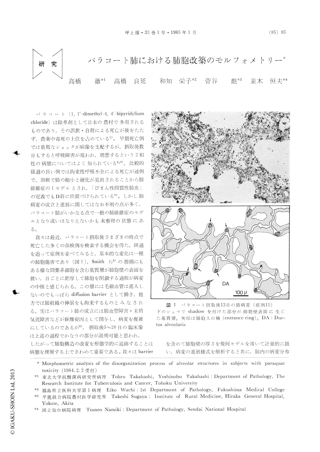
我々は最近,パラコート摂取後さまざまの時点で死亡した多くの剖検例を検索する機会を得た。経過を追って症例を並べてみると,基本的な変化は一種の肺胞傷害であり(図1),Smithら5)の指摘にもある様な間葉系細胞を含む基質層が肺胞壁の表面を被い,日ごとに肥厚して肺胞を閉鎖する過程が病変の中核と感じられる。この層には毛細血管は進入しないのでもっぱらdiffusion barrierとして働き,他方では肺組織の伸展をも拘束するものとみなされる。実はパラコート肺の成立には肺血管障害・末梢気道障害などが修飾要因として関与し,病変を複雑にしているのであるが6),摂取後5〜20日の臨床像は上述の過程でかなりの部分が説明可能と思われ,したがって肺胞構造の改変を形態学的に追跡することは病態を理解する上できわめて重要である。我々はbarrierを含めて肺胞壁の厚さを幾何モデルを用いて計量的に扱い,病変の進展様式を解析すると共に,肺内の病変分布についても検討を行った。
Histopathologic examination was performed of autopsy lungs from fifteen patients who died 20 hours to 23 days after the ingestion of paraquat, disclosing that the basic morbid process consisted in the deposition of matrix substrate which extended on the surface of alveolar septa and in which mesenchymal cells proliferated. Alveolar septa were thickened rapidly and progressively by this deposi-tion, which, working as a diffusion barrier and restricting the distensibility of lung tissues, appearedbasically responsible for inducing severe respiratory failure. In view of this, the mean thickness d of alveolar septa was determined assuming a geometric model, in which the septa in a unit volume of tissue were transformed into a plate that was of a uniform thickness d and had the same surface area Sv as the septa ; Sv was estimated by line sampling on histologic lung sections, applying a basic prin-ciple of stereology. Because the measurement values were influenced more or less by the grade of collapse with which a given lung was fixed, a method to standardize the value of d was devised. The estimates of d remained in the normal range of 10 to 14μ within the first five days after the ingestion of paraquat, while it arose rapidly there-after, reaching 200μ at about the 20th day, whichwas sufficiently thick to obliterate alveolar spaces ; d was shown to follow an exponential function of t. This result coincided well enough with the actual histopathology where, at about 20 days, the shape of ducats and sacculus alveolaris was much sim-plified due to the obliteration of alveoli. On the other hand, in patients who survived relatively long and showed markedly advanced pulmonary changes, the lung never failed to harbor areas where alveoli were maintained uninvolved in fibrotic processes. This was clearly shown in the histogram of d where the variance was much larger in patients of longer survival, corresponding to the marked heterogeneity of pulmonary lesions often experienced in such patients.
Copyright © 1985, Igaku-Shoin Ltd. All rights reserved.