Japanese
English
研究
膜性部中隔動脈瘤の心エコー図所見
Echocardiographic detection of aneurysms of the membraneous septum
豊増 弘幸
1
,
押領司 篤茂
1
,
小須賀 健一
1
,
大石 喜六
1
,
古賀 道弘
1
,
古賀 義則
2
,
陣内 重三
2
,
吉岡 春紀
2
Hiroyuki Toyomasu
1
,
Atsushige Ohryoji
1
,
Ken-ichi Kosuga
1
,
Kiroku Ohishi
1
,
Michihiro Koga
1
,
Yoshinori Koga
2
,
Jyuzo Jinnouchi
2
,
Haruki Yoshioka
2
1久留米大学医学部第2外科
2久留米大学医学部第3内科
1The Second Department of Surgery, Kurume University School of Medicine
2The Third Department of Internal Medicine, Kurume University School of Medicine
pp.761-765
発行日 1979年7月15日
Published Date 1979/7/15
DOI https://doi.org/10.11477/mf.1404203402
- 有料閲覧
- Abstract 文献概要
- 1ページ目 Look Inside
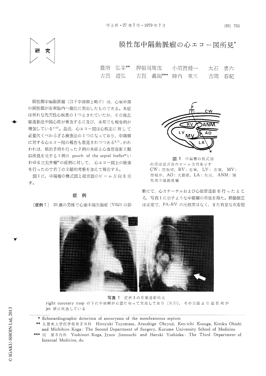
膜性部中隔動脈瘤(以下中隔瘤と略す)は,心室中隔の膜性部が右室腔内へ瘤状に突出したものである。本症は稀れな先天性心疾患の1つとされていたが,その後左室造影法や開心術が普及するに及び,本邦でも報告例が増加している1〜5)。最近,心エコー図は心疾患に対して必要欠くべからざる検査法の1つになっており,中隔瘤に対する心エコー図の報告も散見されつつある6,7)。われわれは,根治手術を行った2例の本症と心血管造影上類似所見を呈する1例のpouch of the septal leaflet8)いわゆる三尖弁瘤9)の症例に対して,心エコー図上の検索を行ったので若干の文献的考察を加えて報告する。
図1に,中隔瘤の模式図と超音波のビーム方向を示す。
Echocardiographic features of two cases with an aneurysm of the membranous septum and a case with a pouch of the septal leaflet were reported.
The diagnosis was made by left ventricular angiography and confirmed by subsequent oper-ation.

Copyright © 1979, Igaku-Shoin Ltd. All rights reserved.


