Japanese
English
- 有料閲覧
- Abstract 文献概要
- 1ページ目 Look Inside
易疲労性,運動時の息切れ,軽度のチアノーゼの他,著しい自覚症状のない28歳の男子で,特異な胸部X線像,心カテーテル所見,心血管造影像などより,肺動脈漏斗部狭窄を伴った完全肺静脈還流異常と診断した1例について報告した。
堂野前維摩郷院長の御校閲を深謝します。
A patient of complete pulmonary venous drainage with infundibular stenosis of the pulmonary ostium is presented. This 28-year-old man has had no serious complaint exceptfor easy fatiguability and occasional breath-lessness, palpitation and light degree of cya-nosis on exertion, since he was checked up cardiac abnormality at the first time in his early childhood. The patient has been well on his ordinary life and job as a welder, and had a baby after he married three years ago.
Physically, his height is 160cm. and wei-ght is 47kg. Cyanosis or clubbed finger is not detectable at rest. Cardiac dullness is widened towards both sides and systolic ej-ection murmur of Levine's grade 3 is audible in the second and third intercostal space of left sternal line. Second pulmonary sound is accentuated and duplicated. Any finding of congestive failure, including hepatomegaly, pulmonary râles and pitting edema on legs, could not be detected.
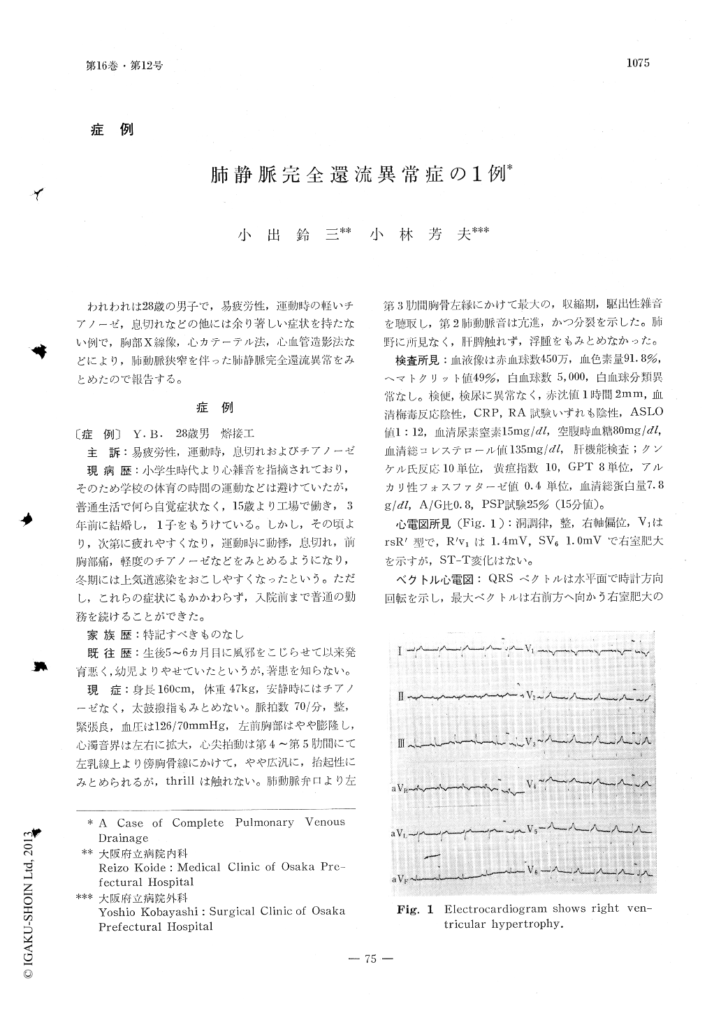
All of the laboratory findings examined are negative, but hematocrit is elevated slightly. Electrocardiogram and vectorcardio-gram reveal right ventricular hypertrophy with abnormal right axis deviation, rsR'pattern in V1 and deep S in V6. On postero-anterior chest X-ray photo, cardiac silhouette shows "the figure of eight" configuration, due to marked distension of upper mediasti-num towards both sides. Pulmonary vascular shadows increase moderately and right ven-tricular hypertrophy is suspected on the films.
Cardiac catheterization reveals elavated right ventricular pressure with normal pul-monary arterial pressure, and oxygen satu-ration is 96 per cent in superior vena cava, 88 per cent in inferior vena cava and 93 per cent in right ventricle, which suggests left to right shunt at some part of superior vena cava. Peripheral arterial oxygen saturation is a little lower than normal.
On angiocardiogram, pulmonary veins of both sides are fused together into common pulmonary vein in back of the heart, which connects to dilatated right superior vena cava, via the remnant left superior vena cava. Another film of the angiocardiogram also shows moderate infundibular stenosis of the pulmonary ostium. Corrected operation was not performed in this case.
The literature on complete pulmonary ven-ous drainage is reviewed, and the possible relationship of reducing pulmonary blood flow by the associated pulmonary stenosis to ra-ther better prognosis in this case, which has no serious signs and symptoms even at the age of twenty eight, is discussed.

Copyright © 1968, Igaku-Shoin Ltd. All rights reserved.


