Japanese
English
- 有料閲覧
- Abstract 文献概要
- 1ページ目 Look Inside
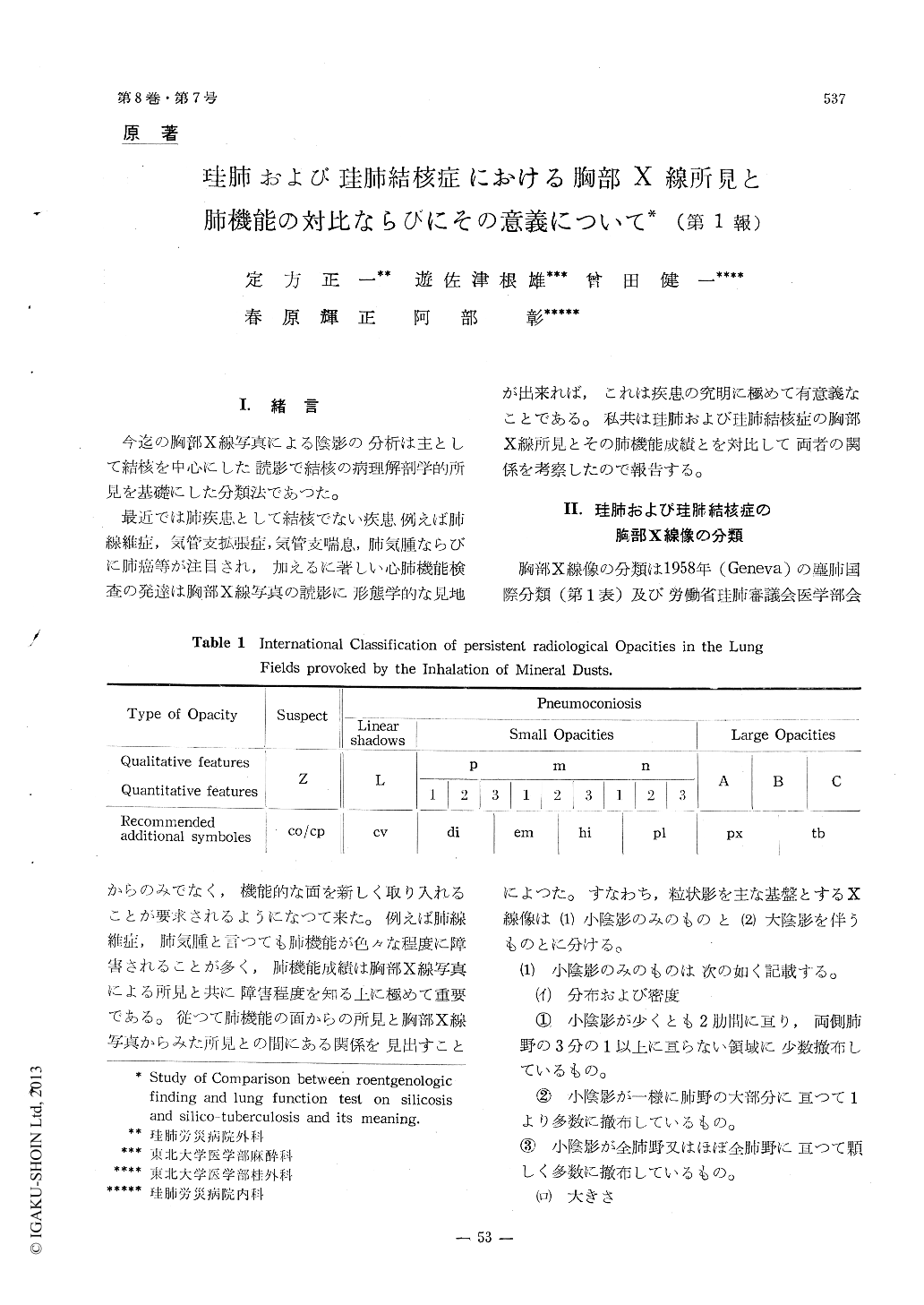
I.緒言 今迄の胸部X線写真による陰影の分析は主として結核を中心にした読影で結核の病理解剖学的所見を基礎にした分類法であつた。
最近では肺疾患として結核でない疾患例えば肺線維症,気管支拡張症,気管支喘息,肺気腫ならびに肺癌等が注目され,加えるに著しい心肺機能検査の発達は胸部X線写真の読影に形態学的な見地からのみでなく,機能的な面を新しく取り入れることが要求されるようになつて来た。例えば肺線維症,肺気腫と言つても肺機能が色々な程度に障害されることが多く,肺機能成績は胸部X線写真による所見と共に障害程度を知る上に極めて重要である。従つて肺機能の面からの所見と胸部X線写真からみた所見との問にある関係を見出すことが出来れば,これは疾患の究明に極めて有意義なことである。私共は珪肺および珪肺結核症の胸部X線所見とその肺機能成績とを対比して両者の関係を考察したので報告する。
Patients which were administered in our hospital were divided into three groups ; 1 st group were patients in whom small opacities were in their x-ray films and 2 nd group were patients in whom small opacities and large opacities were in their x-ray films and thus 3 rd group were patients in whom small opacities, large opacities and tuberculous shadows were in their x-ray films.
In the three groups roentgenologic findings were consisted of symboles that were on the interna-tional classification of persistent radiological opacities in the lung fields provoked by the inhalation of mineral dust (1958, Geneva).
And twelve results of such lung functiontest were calculated on each patients : 1) vital capacity (cc). 2) vital capacity observed volume/predicted volume × 100.3) total lung capacity (cc). 4) timed vital capacity in 1 sec. observed volume/predicted volume ×100.5) residual volume/total lung capacity × 100%.6) Leslie's Index. 7) Index of air trapp-ing. 8) Maximum Breathing capaciy (1). 9) maximum breating capacity ovseved volume/predicted volume ×100.10) air velocity Index. 11) uniformty index. 12) diffusion capacity of the lung (co-sigle breath method)
In consequence of the lung function tests and roentgenologic findings were compared in 1 st group, the lung function was nearly normal in 2 p group but in 3 m group loss of lung function was much and more in 3 p group. Moreover patints only diffusion capacities of the lungs were lower were observed.

Copyright © 1960, Igaku-Shoin Ltd. All rights reserved.


