Japanese
English
- 有料閲覧
- Abstract 文献概要
- 1ページ目 Look Inside
- 参考文献 Reference
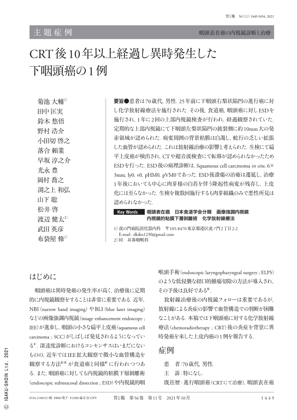
要旨●患者は70歳代,男性.25年前に下咽頭右梨状陥凹の進行癌に対し化学放射線療法を施行された.その後,食道癌,咽頭癌に対しESDを施行され,1年に2回の上部内視鏡検査が行われ,経過観察されていた.定期的な上部内視鏡にて下咽頭左梨状陥凹の披裂側に約10mm大の発赤領域が認められた.病変周囲の背景粘膜は白濁し,蛇行の乏しい拡張した血管が認められた.これは放射線治療の影響と考えられた.生検にて扁平上皮癌が検出され,CTや超音波検査にて転移が認められなかったためESDを行った.ESD後の病理診断は,Squamous cell carcinoma in situ,6×3mm,ly0,v0,pHM0,pVM0であった.ESD後潰瘍の治癒は遷延し,治療1年後においても中心に肉芽様の白苔を伴う隆起性病変が残存し,上皮化には至らなかった.生検を複数回施行するも肉芽組織のみで悪性所見は認められなかった.
The patient was a man in his 70s. Twenty-five years ago, he underwent chemoradiotherapy for advanced cancer of the right pyriform sinus of the hypopharynx. This was followed by ESD(endoscopic submucosal dissection)for esophageal cancer and pharyngeal cancer and then upper gastrointestinal endoscopy twice a year for follow-up.
A reddish area approximately 10mm in size was detected in the left pyriform sinus of the hypopharynx. Irregular microvessels without loop formation were detected, which were classified as B2 vessels according to the Japan Esophageal Society's classification. Biopsy revealed squamous cell carcinoma, and computed tomography and ultrasound showed no lymph node or distant metastases. Our final diagnosis was superficial pharyngeal carcinoma with subepithelial invasion. ESD was performed under general anesthesia, and the lesion was resected en bloc. The pathological result after ESD was squamous cell carcinoma in situ, 6×3mm, ly0, v0, margin negative. Healing of the ulcer after ESD was prolonged. One year after the treatment, a raised lesion with granulomatous white moss remained in the center ; however, epithelialization did not occur.
Copyright © 2021, Igaku-Shoin Ltd. All rights reserved.