Japanese
English
- 有料閲覧
- Abstract 文献概要
- 1ページ目 Look Inside
- 参考文献 Reference
- サイト内被引用 Cited by
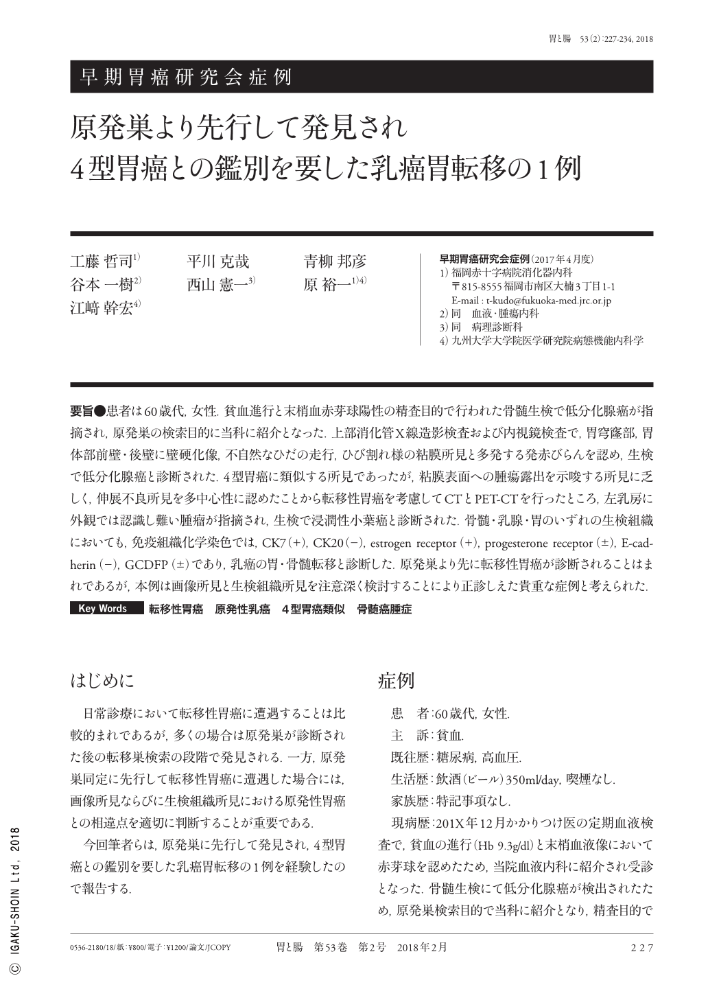
要旨●患者は60歳代,女性.貧血進行と末梢血赤芽球陽性の精査目的で行われた骨髄生検で低分化腺癌が指摘され,原発巣の検索目的に当科に紹介となった.上部消化管X線造影検査および内視鏡検査で,胃穹窿部,胃体部前壁・後壁に壁硬化像,不自然なひだの走行,ひび割れ様の粘膜所見と多発する発赤びらんを認め,生検で低分化腺癌と診断された.4型胃癌に類似する所見であったが,粘膜表面への腫瘍露出を示唆する所見に乏しく,伸展不良所見を多中心性に認めたことから転移性胃癌を考慮してCTとPET-CTを行ったところ,左乳房に外観では認識し難い腫瘤が指摘され,生検で浸潤性小葉癌と診断された.骨髄・乳腺・胃のいずれの生検組織においても,免疫組織化学染色では,CK7(+),CK20(−),estrogen receptor(+),progesterone receptor(±),E-cadherin(−),GCDFP(±)であり,乳癌の胃・骨髄転移と診断した.原発巣より先に転移性胃癌が診断されることはまれであるが,本例は画像所見と生検組織所見を注意深く検討することにより正診しえた貴重な症例と考えられた.
A 69-year-old female was referred to our institution because of anemia with erythroblasts. Bone marrow biopsy revealed bone metastasis, which was histopathologically diagnosed as poorly differentiated adenocarcinoma. Radiography revealed multifocal scirrhous lesions. On performing EGD(esophagogastroduodenoscopy), thickening of the gastric folds, resembling primary type IV gastric cancer, was observed. Because no superficial lesion of primary gastric cancer was observed throughout the gastric wall, we suspected these gastric lesions to be metastatic cancer. CT and PET-CT demonstrated a left mammary tumor. Endoscopic biopsy revealed poorly differentiated adenocarcinoma with signet ring cells. Core needle biopsy of the breast tumor showed invasive lobular carcinoma. In the bone marrow, stomach, and mammary gland, all the tumor cells were immunohistochemically positive for estrogen receptor, progesterone receptor, GCDFP, and CK7 but negative for CK20 and E-cadherin. Based on these findings, the patient was diagnosed with primary breast cancer that metastasized to the stomach and bone marrow. This is a rare case because the diagnosis of metastatic gastric cancer preceded the detection of primary breast cancer. Therefore, careful observation of radiographic, endoscopic, and pathologic findings is important for an accurate diagnosis.

Copyright © 2018, Igaku-Shoin Ltd. All rights reserved.


