Japanese
English
- 有料閲覧
- Abstract 文献概要
- 1ページ目 Look Inside
- 参考文献 Reference
- サイト内被引用 Cited by
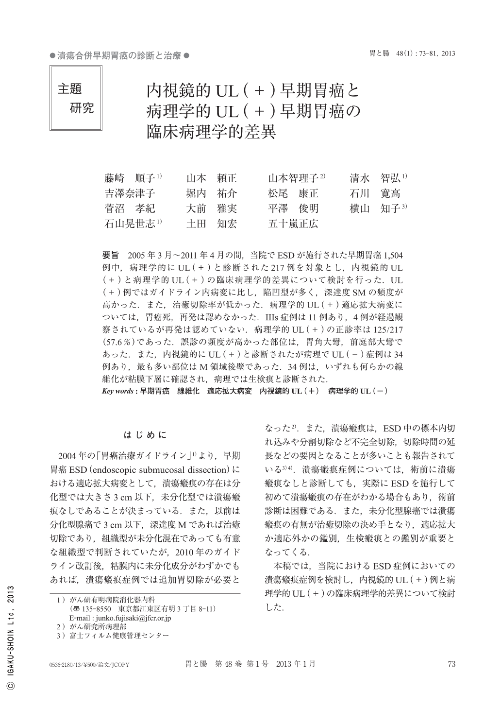
要旨 2005年3月~2011年4月の間,当院でESDが施行された早期胃癌1,504例中,病理学的にUL(+)と診断された217例を対象とし,内視鏡的UL(+)と病理学的UL(+)の臨床病理学的差異について検討を行った.UL(+)例ではガイドライン内病変に比し,陥凹型が多く,深達度SMの頻度が高かった.また,治癒切除率が低かった.病理学的UL(+)適応拡大病変については,胃癌死,再発は認めなかった.IIIs症例は11例あり,4例が経過観察されているが再発は認めていない.病理学的UL(+)の正診率は125/217(57.6%)であった.誤診の頻度が高かった部位は,胃角大彎,前庭部大彎であった.また,内視鏡的にUL(+)と診断されたが病理でUL(-)症例は34例あり,最も多い部位はM領域後壁であった.34例は,いずれも何らかの線維化が粘膜下層に確認され,病理では生検痕と診断された.
In this study, we investigated the clinicopathological differences between gastric cancer patients with endoscopic ulcer scars and pathological ulcer scars. Towards this end, we selected 217 gastric cancer patients who were confirmed histopathologically as having ulcer scars, from among 1,504 patients who had undergone ESD(endoscopic submucosal dissection)at our hospital between 2005 and April 2011. Among the patients with the pathological ulcer scars(+), cases with the depressed type of cancer were many, and the percentage of cases classified, according to the depth of invasion, as SM was higher in comparison to the lesions described in the guidelines. In addition, the rate of curative resection in this group was also lower. For cases of pathological ulcer scars(+)not falling within the description provided in the guidelines, there were neither cases of death from gastric cancer nor cases with recurrence of cancer. There were 11 cases of stage III, of which 4 were followed up, but were not detected to have any recurrence. The accurate endoscopic diagnosis rate of pathological ulcer scars(+)was 92/217(57.6%). Regions of high incidence of inaccurate diagnosis were the greater curvature at the angular incisure and the greater curvature at the gastric antrum. There were 34 patients who were diagnosed as pathological ulcer scar(-)despite being diagnosed as endoscopic ulcer scar(+), and the most frequent region of detection of the false-positive scars was the posterior wall of the M region. In all of these 34 cases, some fibrosis was observed in the submucosa, and the diagnosis of biopsy scar was confirmed by pathological examination.

Copyright © 2013, Igaku-Shoin Ltd. All rights reserved.


