Japanese
English
- 有料閲覧
- Abstract 文献概要
- 1ページ目 Look Inside
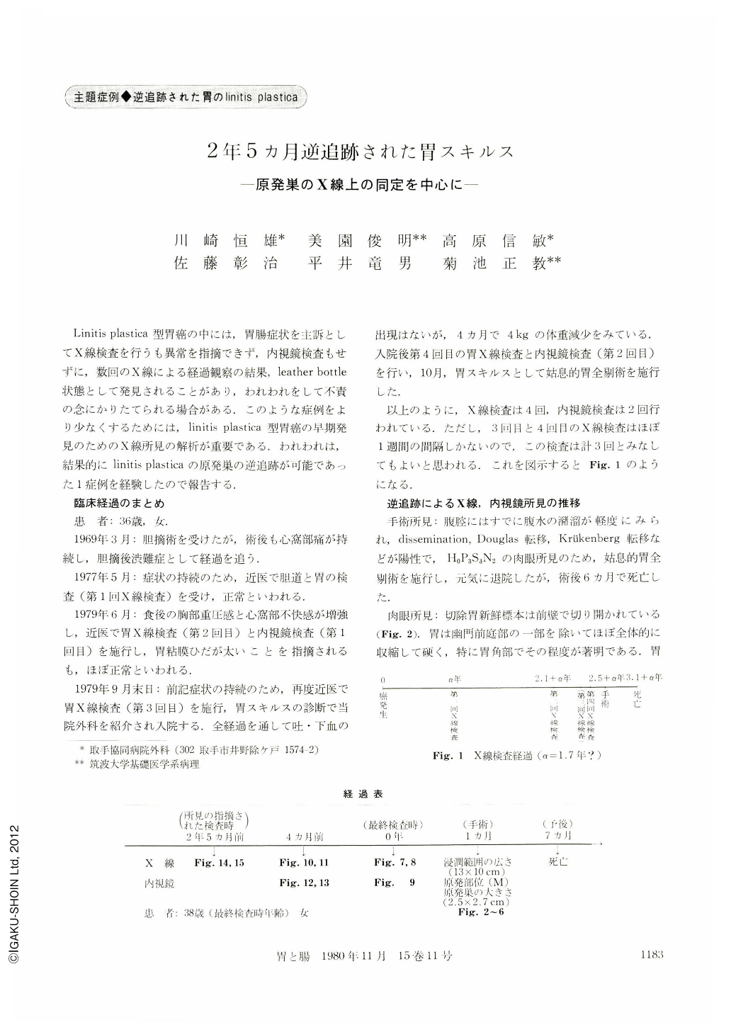
Linitis plastica型胃癌の中には,胃腸症状を主訴としてX線検査を行うも異常を指摘できず,内視鏡検査もせずに,数回のX線による経過観察の結果,leather bottle状態として発見されることがあり,われわれをして不責の念にかりたてられる場合がある.このような症例をより少なくするためには,linitis plastica型胃癌の早期発見のためのX線所見の解析が重要である.われわれは,結果的にlinitis plasticaの原発巣の逆追跡が可能であった1症例を経験したので報告する.
A 36 year-old woman had been diagnosed as having gastric carcinoma of linitis plastica type (typical leather bottle) by an x-ray examination of the upper GI-tract (Fig. 7, 8) and total gastrectomy had been carried out at our hospital. Histological examination of resected stomach revealed a primary focus of the carcinoma, located at the posterior wall of the corpus near the greater curvature and measuring approximately 2.7×2.5 cm in dimensions (Fig. 2, 3). Furthermore, the primary focus was limited to the fundic gland mucosa area without intestinal metaplasia, and the oral three-quarters of the gastric wall were infiltrated diffusely with cancer cells with marked desmoplasia in the gastric layers except the mucosa.
She had had an x-ray examination of the upper GI-tract in 2 years and 5 months before the surgery at one out-patient clinic. Although the x-ray pictures firstly examined were not so good for diagnosis, they have been studied retrospectively from the view-point of a hypothesis of growing process of carcinoma of linitis plastica type in cancer development. It is evident from the histological findings of the resected stomach that the primary focus was situated at the posterior wall of the corpus near the greater curvature, which showed macroscopically the area with many mucosal rugae. According to the hypothesis, the primary focus may be shown radiologically as type Ⅱc without convergency of the mucosal rugae, and the size could be estimated as approximately 1 cm in the largest diameter. Consequently, we have been able to determine the primary focus as type Ⅱc without convergency of mucosal rugae and measuring around 1 cm in the largest diameter (Fig. 15).

Copyright © 1980, Igaku-Shoin Ltd. All rights reserved.


