Japanese
English
- 有料閲覧
- Abstract 文献概要
- 1ページ目 Look Inside
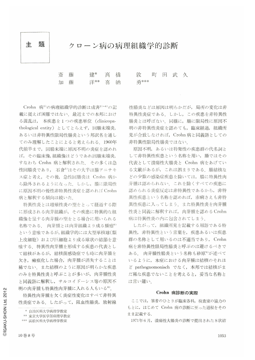
Crohn病1)の病理組織学的診断は成書2)~4)の記載に従えば困難ではない.最近までの本邦における混乱は,本疾患を1つの疾患単位(clinicopathological entity)としてとらえず,回腸末端炎,あるいは非特異性限局性腸炎という邦訳名を通してのみ理解したことによると考えられる.1960年代前半まで,回腸末端に原因不明の炎症を認めれば,その臨床像,組織像はどうであれ回腸末端炎,すなわちCrohn病と解釈された.その多くは急性回腸炎であり,石倉5)はその大半は腸アニサキス症と考え,その後,急性回腸炎はCrohn病から除外されるようになった.しかし,腸に限局性に原因不明の慢性非特異性炎症を認めればCrohn病と解釈する傾向は続いた.
特異性炎とは増殖性炎の型をとって経過する際に形成される肉芽組織が,その疾患に特異的な組織像を呈する肉芽腫の型をとる場合に用いられる名称である.肉芽腫とは肉芽組織より成る腫瘤6)という意味であるが,組織学的には大型単核球(類上皮細胞)および巨細胞より成る球状の結節を意味する.特異性肉芽腫を形成する疾患の代表として結核があるが,結核菌感染症でも時に肉芽腫を欠き,瘢痕化した場合,肉芽腫が消失することは稀でない.また結核のように原因が明らかな疾患のみを特異性炎と呼ぶことが多いが,肉芽腫性炎と同義語に解釈し,サルコイドーシス等の原因不明の肉芽腫も特異性肉芽腫に入れる人もいる8).
All of the resected intestinal segments excluding duodenum, examined at the Section of Pathology, Tokyo University Hospital from 1955 to 1974, Hitachi General Hospital from 1960 to 1974 and Jichi Medical School Hospital in 1974 were reviewed.
There were only 12 cases of Crohn's disease characterised by transmural inflammation, fissuring ulcers and non-caseating epitheloid cell granulomata. Ten cases were male and 2 cases were female. The age of the patients at the time of operation was from 16 years old to 34 years (average 25 years). Sites of the lesions were as follow: Terminal ileum, 3 cases; terminal ileum, caecum and ascending colon, 2 cases; caecum, 1 case; ascending colon, 3 cases; entire colon, 1 case; rectum, 1 case; terminal ileum, entire colon, and duodenum, 1 case, which was the first case of histologically confirmed duodenal Crohn's disease in Japan. Epitheloid cell granuloma was identified in 10 cases. Appendiceal lesion was ascertained in 3 cases. Anal lesion was described in 2 cases. Intestinal Behçet had been suspected in a case of rectal Crohn's disease because of aphthoid lesions of the mouth and perianal ulcer.
Most cases diagnosed as ulcerative colitis have been treated by medications. Nineteen cases were surgically treated in the same period. Nine cases were male and 10 cases were female. The age of the patients at the time of first operation was from 18 years to 45 years (average 27 years).
Thirty four cases of intestinal tuberculosis were resected in the same period. Of these, 16 cases had ileocaecal lesions, 12 cases had lesions in small intestine other than terminal ileum. Six cases had colonic lesions. The average age at the time of resection was 36 years.
Special interest was paid to solitary, large, deep ulcer of the caecun involing the Bauhin's valve, often called as “non-specific inflammatory tumor of the colon” or “non-specific ulcer of the colon” in Japan. The large ulcer with deep undermining was usually located in the mesenteric side of the caecum and penetration of the entire wall was the rule. Follicular lymphocytic aggregations were often found around the ulcer. The entire intestinal wall near the ulcer was normal and no epitheloid cell granulomata was discovered. The average age of the patients was 27 years. Two cases over the age of 50 were male and we could not completely exclude postappendectomy complication. Twelve cases were under the age of 35, of which 3 cases were male and 9 cases were female. Recurrence of ulcer at the site of anastomosis was documented in 2 cases.
No case consistent “ulcerative, non-proliferative disease of the small intestine (Okabe, Sakimura)” was found in our materials. Three cases over the age of 50 revealed multiple ulcer scars of the small intestine which resembled healed intestinal tuberculosis although no evidence was identified as such at the time of operation.

Copyright © 1975, Igaku-Shoin Ltd. All rights reserved.


