Japanese
English
- 有料閲覧
- Abstract 文献概要
- 1ページ目 Look Inside
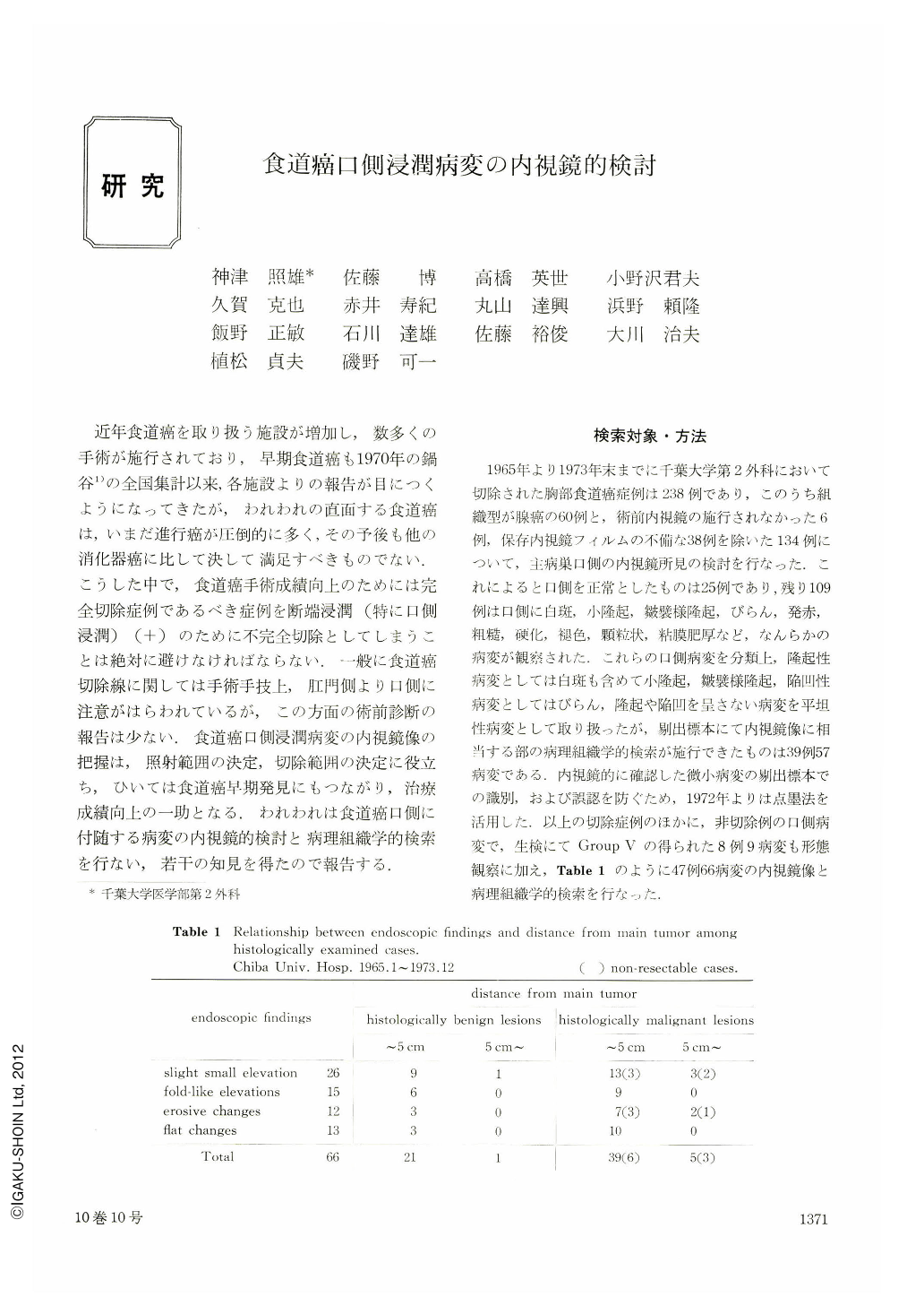
近年食道癌を取り扱う施設が増加し,数多くの手術が施行されており,早期食道癌も1970年の鍋谷1)の全国集計以来,各施設よりの報告が目につくようになってきたが,われわれの直面する食道癌は,いまだ進行癌が圧倒的に多く,その予後も他の消化器癌に比して決して満足すべきものでない.こうした中で,食道癌手術成績向上のためには完全切除症例であるべき症例を断端浸潤(特に口側浸潤)(+)のために不完全切除としてしまうことは絶対に避けなければならない.一般に食道癌切除線に関しては手術手技上,肛門側より口側に注意がはらわれているが,この方面の術前診断の報告は少ない.食道癌口側浸潤病変の内視鏡像の把握は,照射範囲の決定,切除範囲の決定に役立ち,ひいては食道癌早期発見にもつながり,治療成績向上の一助となる.われわれは食道癌口側に付随する病変の内視鏡的検討と病理組織学的検索を行ない,若干の知見を得たので報告する.
Precise endoscopic confirmation of oral side invasion of esophageal carcinoma is very useful for deciding the resective line and irradiation field, and moreover is one of the important measures for finding out early carcinoma and for promoting therapeutic results.
We classified the oral side changes accompanying esophageal carcinoma into slight small elevation, fold-like elevation, erosion and flat changes, such as reddening, roughness, discoloration and hardening.
We studied endoscopically and histologically the oral side changes of 134 cases of thoracic esophageal carcinoma in our 9 years' experience, excluding adenocarcinoma in histological type. For confirming the lesions, we used the pre-operative mucosal tattooing.
Slight small elevations, proven to be malignant histologically, are mostly yellow or not different from surrouuding tissues in color, and are in size over rice grain and have ill-defined margin. When the carcinoma is exposed over the epithelial layer, the elevation becomes white and its margin becomes welldefined. We experienced a lesion of intramural metastasis 9 cm orally apart from the maintumor.
Malignant fold-like elevation has a shift of the origin over to the oral side of the main tumor center. When the origin is situated on the margin, it has yellow elevation on a fold or a widening of folds.
Malignant erosions are mostly observed already before pre-operative irradiation, and they are welldefined in margin and slightly or moderately reddened and are observed in a lesion with wide area. They are often observed at some distance from the main tumor, in one case even 6 cm orally.
Flat lesions are not usually observed singly. They rea seen as reddening in carcinoma in situ and as discoloration or hardening in subepithelial cancer infiltration. Preoperative confirmation of submucosal cancer infiltration reems to be future problems for endoscopic examination.

Copyright © 1975, Igaku-Shoin Ltd. All rights reserved.


