Japanese
English
- 有料閲覧
- Abstract 文献概要
- 1ページ目 Look Inside
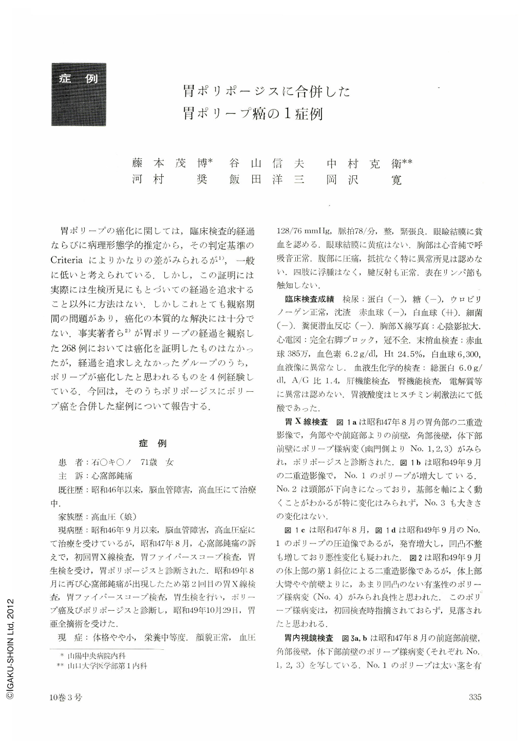
胃ポリープの癌化に関しては,臨床検査的経過ならびに病理形態学的推定から,その判定基準のCriteriaによりかなりの差がみられるが,一般に低いと考えられている.しかし,この証明には実際には生検所見にもとづいての経過を追求すること以外に方法はない.しかしこれとても観察期間の問題があり,癌化の本質的な解決には十分でない.事実著者らが胃ポリープの経過を観察した268例においては癌化を証明したものはなかったが,経過を追求しえなかったグループのうち,ポリープが癌化したと思われるものを4例経験している.今回は,そのうちポリポージスにポリープ癌を合併した症例について報告する.
Cancerous change of gastric polyp is of very infrequent occurence, but its possibility should always be borne in mind, because polyp cancer does exist. In the past we have come across 4 cases of the so-called polyp cancer of the stomach associated with gastric polyposis.
The patient, a 71-year-old woman, had been diagnosed as harboring gastric polyposis 2 years and a month before when she had undergone minute examination of the stomach, including x-ray, endoscopy and biopsy, on account of pain in the epigastrium. Recently she was re-examination by x-ray and endoscopy because of the same complaint, A big polyp (No. 1), enlarged since the initial examination, was seen on the posterior wall at the level of the angle in the neighborhood of the antrum, with two polyps (No. 2 and 3) each on the posterior wall of the angle and on the anterior wall of the lower body, both of the same size as before, and another polypoid lesion in the side of the anterior wall on the greater curvature of the upper body (No. 4, initially overlooked ?). They were all pedunculated. Polyp No. 1 had grown up to such a size that cancerous change could not be ruled out.
Unlike others, polyp No. 4 was entirely covered with pale whitish coat. By biopsy it was shown to belong to group Ⅳ. Other polyps were of adenomatos nature. Gross specimen of the resected stomach showed that polyp No. 1 was bigger than others, measuring 2.6cm in the largest diameter at the tip, markedly uneven on the surface with reddish color. Grossly too, it was impossible to rule out malignancy. Polyp No. 4 was also of tall stature, 2.4cm in height, and of more blackish tone than the others. It was likewise impossible to tell whether or not it was cancerous. Histologically, tubular adenocarcinoma was recognized in a portion adjacent to the tip of the polyp No. 4. The remainders were all of adenomatous nature.
The present case is of considerable interest because a polyp cancer was seen side with another larger polyp that had been observed to grow up and increase in size.

Copyright © 1975, Igaku-Shoin Ltd. All rights reserved.


