Japanese
English
- 有料閲覧
PPV購入案内
1,320円 (1,200円+税10%) こちらは、595ページから600ページの文献です。購入には医書.jp本会員登録が必要です。
会員登録完了後に改めて上記「購入サイトへ」を選択してください。
または「購入サイトへ」選択後に「購入ログイン」、「新規会員登録」の順に進んでください。
会員登録について詳しくはをご確認ください。
- Abstract 文献概要
- 1ページ目 Look Inside
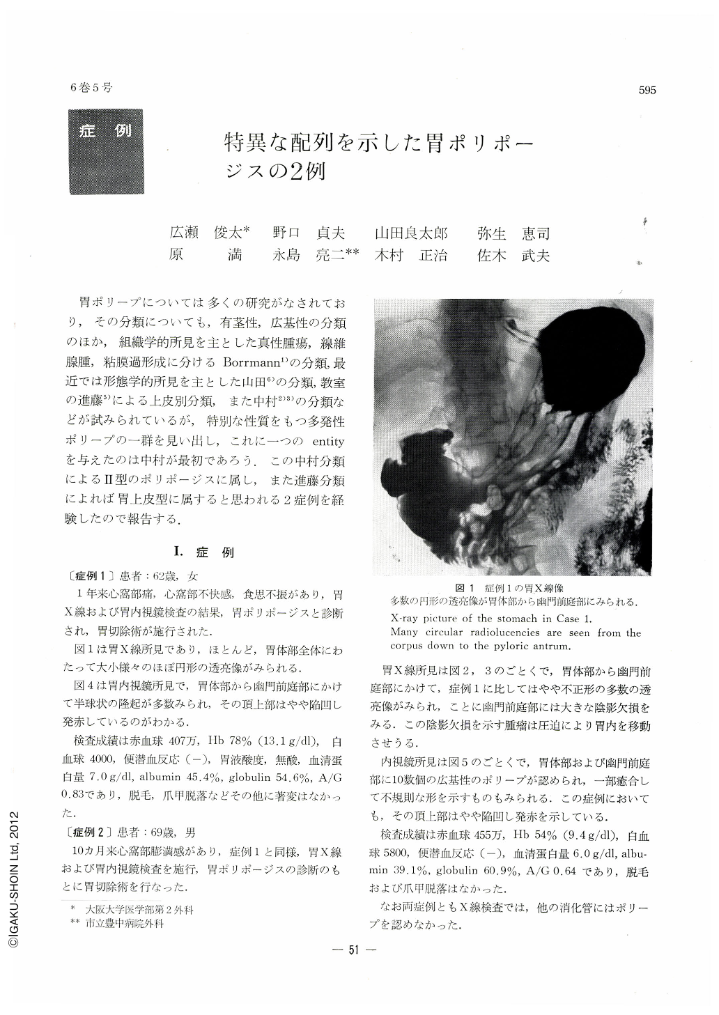
胃ポリープについては多くの研究がなされており,その分類についても,有茎性,広基性の分類のほか,組織学的所見を主とした真性腫瘍,線維腺腫,粘膜過形成に分けるBorrmann1)の分類,最近では形態学的所見を主とした山田6)の分類,教室の進藤5)による上皮別分類,また中村2)3)の分類などが試みられているが,特別な性質をもつ多発性ポリープの一群を見い出し,これに一つのentityを与えたのは中村が最初であろう.この中村分類によるⅡ型のポリポージスに属し,また進藤分類によれば胃上皮型に属すると思われる2症例を経験したので報告する.
Gastric polyp was classified by Nakamura (1966) into four types, The second type was defined as following: multiple polyps developing in a part of the corpus adjacent to the pyloric gland region and having tips with engorged dimples. This variety of polyp is considered to have grown by the repetition of erosion and repair followed by reactive hyperplasia of the surrounding mucosa.
In this paper are described two cases of this type of gastric polyposis recently experienced.
Cases: Both Case 1 (a 62-year-old woman) and Case 2 (a 69-year-old man) had complained of pain and sensation of fullnes in the epigastrium. X-ray and endoscopic examinations of the stomach revealed in both cases many sessile polyps in the corpus and antrum. In the latter, a large filling defect was also recognized in the antrum. Accordingly, they were operated on under the diagnosis of gastric polyposis.
Gross view of the resected specimens: Polyps were distributed in a V-like fashion along the border between the corpus (fundic gland region) and antrum (pyloric gland region), measuring 5 to 10 mm in diameter, round and sessile, with engorged dimples at the tip. In the second case, a large polyp that seemed to be fusion of several polyps was seen besides smaller ones on the posterior wall of the antrum.
Histological findings: In both instances the muscularis mucosae was generally thick at the bases of all the polyps, some protruding well into the growths. It was clear that, though grossly sessile, these polyps had stalks histologically. Their central dimples were slightly eroded. A large part of the glandular epithelium were foveolar and the border region between the glands consisted of pseudopyloric glands and cysts. Epithelial cells of the polyps were neither atypical nor malignant.
Both macroscopically and microscopically, the polyps in the present cases are considered to belong to the second type as classified by Nakamura.
Copyright © 1971, Igaku-Shoin Ltd. All rights reserved.