Japanese
English
- 有料閲覧
- Abstract 文献概要
- 1ページ目 Look Inside
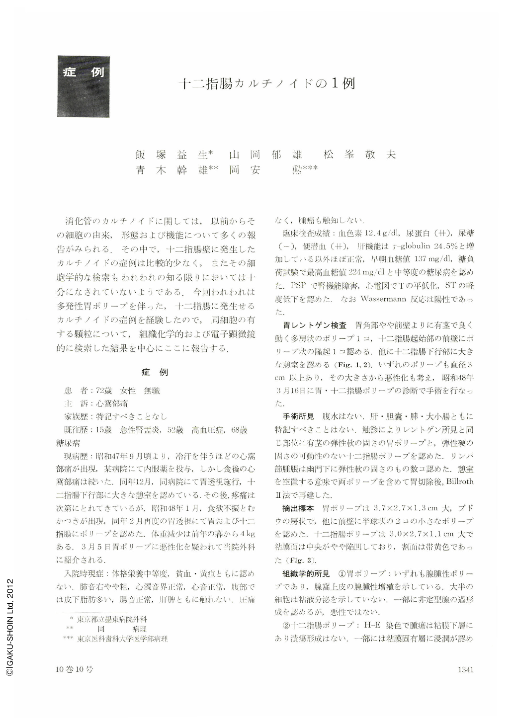
消化管のカルチノイドに関しては,以前からその細胞の由来,形態および機能について多くの報告がみられる.その中で,十二指腸壁に発生したカルチノイドの症例は比較的少なく,またその細胞学的な検索もわれわれの知る限りにおいては十分になされていないようである.今回われわれは多発性胃ポリープを伴った,十二指腸に発生せるカルチノイドの症例を経験したので,同細胞の有する顆粒について,組織化学的および電子顕微鏡的に検索した結果を中心にここに報告する.
A 72-year-old woman visited our hospital with a chief complaint of epigastralgia in March 1973. There was no carcinoid syndrome in her history. She was diagnosed to harbor gastric and duodenal polyps by upper gastrointestinal roentgenograms. Gastrectomy was performed then.
Gross specimen of the resected stomach showed a large polyp, 3.7×2.7×1.3 cm, on the anterior wall of the midbody, with two small polyps near it, and another large polypoid lesion, 3.0×2.7×1.1 cm, on the anterior wall of the duodenum. The latter was a submucosal tumor, indented on the surface and its cut surface showing yellow color, so that it was diagnosed as carcinoid tumor of the duodenum. Histologically tumor cells of uniform size showed cord-like, island-like or sheet-like structure.
Argyrophil reaction by Grimelius method showed argylophil granules, but argentaffine reaction by Masson-Fontana method was negative. These reaction are typical of foregut origin carcinoids.
Electromicroscopy showed small rounded cells containing round nuclei with carcinoid granules in the cytoplasm. These granules were of two sizes, the smaller ones measuring 130~200 mμ and larger ones 340~470 mμ. They were of dark round form, encased by dense single membrane. Microvilli were not seen. Serotonin and 5-HIAA examinations were not performed.

Copyright © 1975, Igaku-Shoin Ltd. All rights reserved.


