Japanese
English
- 有料閲覧
- Abstract 文献概要
- 1ページ目 Look Inside
十二指腸良性腫瘍は比較的稀な疾患とされていたが,近年の消化器診断技術の発達と胃集検の普及とによりその増告が増加してきた,著者らのひとり中村ら1)(1969年)が集計した当時は欧米で422例,本邦で87例であったが,その後の本邦報告例をみると200例近くになる.
著者らの症例も十二指腸良性腫瘍は経過観察例15例,手術例5例,計20例におよんでいる.手術例のうち乳頭状腺腫は1例であり,比較的稀な症例と考えられるので報告する.あわせて文献的考察を加える.
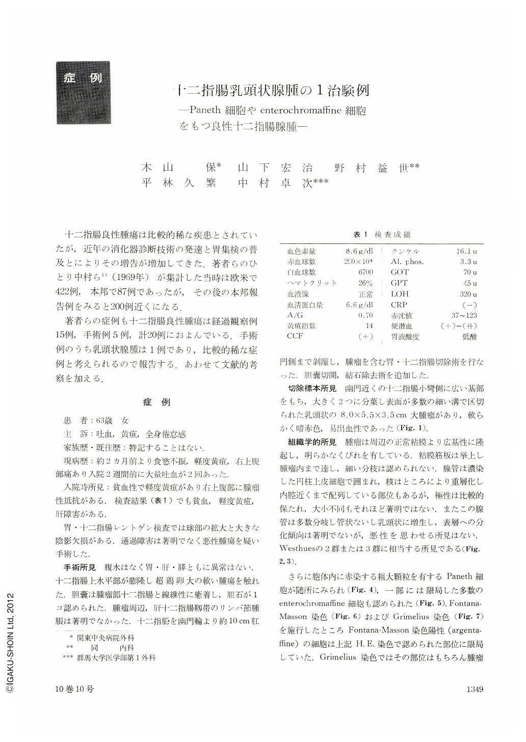
The patient, a 63-year-old woman, was admitted to our hospital on account of pain in the RUQ, hematemesis and jaundice. At admission anemia and a tumor-like mass in the RUQ were noticed. X-ray examination of the upper digestive tract revealed dilated duodenal bulb with a large shadow defect within it. Passage of contrast medium was not hindered. Under a suspicion of malignancy surgical intervention was done, only to find a soft tumor more than the size of an egg in the first limb of the duodenum which was distended. Gastroduodenectomy was accordingly performed. In addition, a gallstone was removed.
The resected specimen showed a broad-based papillary tumor, 8.0×5.5×3.5 cm in dimensions, located in the side of the lesser curvature of the bulb near the pylorus. It was of soft consistency, dark red and easy to bleed.
Histologic study showed that glandular tubules consisted of deep-staining columnar epithels. The nuclei were stratified, but their polarity was relatively well preserved with no too great difference in their size. Glandular tubules sprouted a great many branches, showing tubular or papillary hyperplasia. No differentiating tendency was seen toward the superficial layer, nor was there any finding suggesting malignancy. The whole picture corresponded to Westhue's Group 2 or 3. Numerous Paneth's cells with red-staining rough granules within the cytoplasm were also seen. In some parts were recognized enterochromaffine cells too.
These findings led us to a diagnosis of benign papillary adenoma of the duodenum. The postoperative course was uneventful. The patient died seven years later of other causes unknown.
Adenoma of the digestive tract is usually most often seen in the large intestine. For that of the duodenum we have only reports of 35 cases (26 in segments other than the papilla and 9 in the papillary region). Huge adenoma such as this is reportedly seen only in 4 cases. Furthermore, detailed histological findings such as its atypicality, the presence or absence of Paneth's cells are so scarce that we deemed this case worth reporting with reference to its literature.
Copyright © 1975, Igaku-Shoin Ltd. All rights reserved.