Japanese
English
- 有料閲覧
- Abstract 文献概要
- 1ページ目 Look Inside
N-methyl-N'-nitro-N-nitrosoguanidine(MNNG)の経口投与により,ラットに高率に胃癌を作ることが可能となったことは,杉村らの報告にもあるように1)~3),胃癌の実験的研究に有力な材料を提供した.MNNGは強力な突然変異誘起物質であり4),その発癌性の証明はinvivo5)~7)およびin vitro8)においてなされている.
実験胃癌の組織発生に関する研究は,MNNGの開発以前においても,その胃癌誘発率の低頻度にもめげず行なわれてきた.Howes and De oliveiraは,20-Methylcholanthrene(MC)に浸した糸をラット腺胃の粘膜と漿膜の間に縫い込み,腺胃における初期変化について観察した9).Grant and Ivyは,ほぼ同様の実験操作を行なったラットで,胃粘膜の異常な変化を詳細に報告した10).Stewart一派は,胃癌の判定規準を提唱し,precancerとcancerを区別したが11),さらにMCをマウスの腺胃に注射してその経過を追求し,胃癌は多発性に発生すること,初発巣はumbilicate lesion下の粘膜深層から起こることなどを示唆した12).次いでN,N'-2,7-fluorenylenevisacetamideの経口投与による胃癌発生を報告した13).
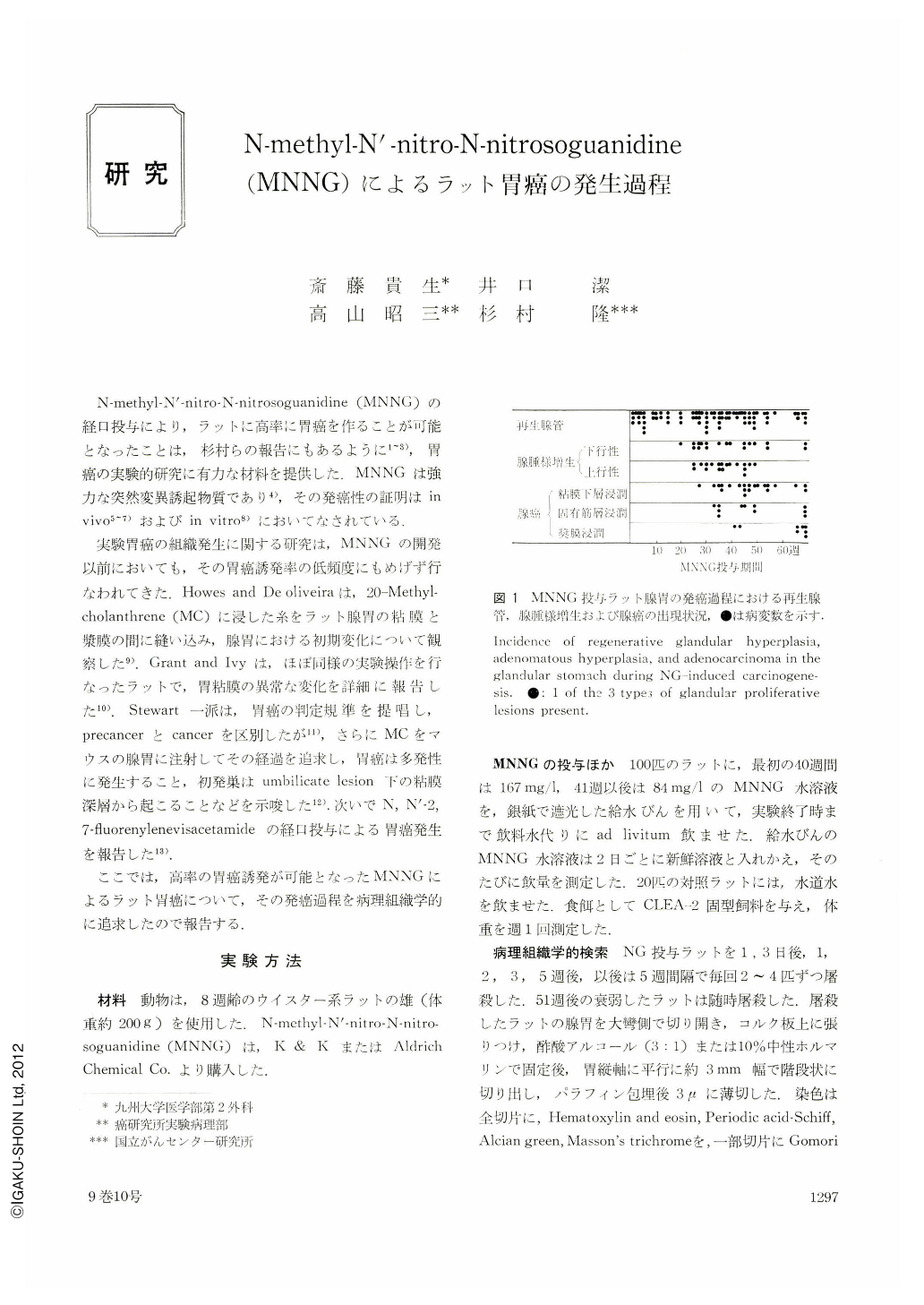
Morphological changes were studied in the glandular stomach of rats with carcinogenesis induced by N-methyl-N'-nitro-N-nitrosoguanidine (MNNG). MN-NG was given to 100 rats in their drinking water at the level of 167 mg/1 for 40 weeks and then 84 mg/1 until the end of the experiment. Rats were killed periodically and histological changes in the glandular stomach were examined. Three types of glandular proliferation were distinguished: regenerative glandular hyperplasia, adenomatous hyperplasia, and adenocarcinoma. These lesions were mainly in the antral region of the stomach. After atrophy and erosion of the mucosa, regenerative glandular hyperplasia, with irregular glands at the margins of erosions, developed in the 3rd-5th week, and these lesions persisted for the rest of the experiment. Adenomatous hyperplasia, characterized by excessive glandular proliferation with scanty cellular atypism, developed in the 20th week, and these lesions were found in the middle of the experimental period. Adenocarcinoma, consisting of excessive glandular proliferation with cellular atypism, appeared in the 30th week and gradually invaded to the submucosa, the muscularis propria, and the serosa. After the 60th week, almost all rats had adenocarcinoma with serosal invasion.

Copyright © 1974, Igaku-Shoin Ltd. All rights reserved.


