Japanese
English
- 有料閲覧
- Abstract 文献概要
- 1ページ目 Look Inside
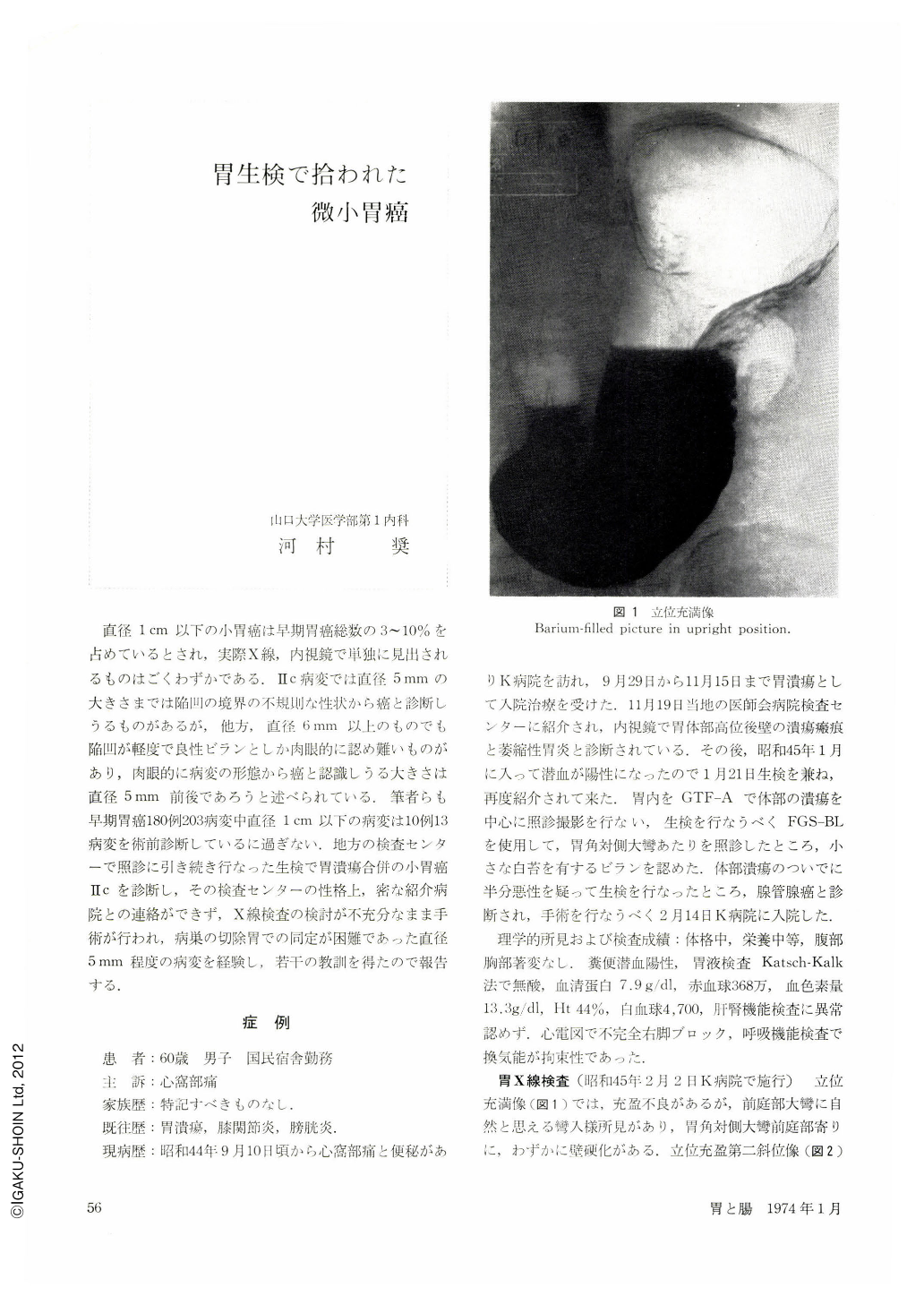
直径1cm以下の小胃癌は早期胃癌総数の3~10%を占めているとされ,実際X線,内視鏡で単独に見出されるものはごくわずかである.Ⅱc病変では直径5mmの大きさまでは陥凹の境界の不規則な性状から癌と診断しうるものがあるが,他方,直径6mm以上のものでも陥凹が軽度で良性ビランとしか肉眼的に認め難いものがあり,肉眼的に病変の形態から癌と認識しうる大きさは直径5mm前後であろうと述べられている.筆者らも早期胃癌180例203病変中直径1cm以下の病変は10例13病変を術前診断しているに過ぎない.地方の検査センターで照診に引き続き行なった生検で胃潰瘍合併の小胃癌Ⅱcを診断し,その検査センターの性格上,密な紹介病院との連絡ができず,X線検査の検討が不充分なまま手術が行われ,病巣の切除胃での同定が困難であった直径5mm程度の病変を経験し,若干の教訓を得たので報告する.
Minute carcinoma of the stomach under 1 cm in diameter reportedly accounts for 3 to 10 per cent of all early gastric cancer. Our results have shown a similar rate. Of 180 cases with 203 lesions of early gastric cancer encountered so far, accurate preoperative diagnosis was made in only 10 cases with 13 lesions.
The case here presented concerns a 60-year-old man who had once been admitted to the hospital on account of gastric ulcer on the posterior wall of the part of the corpus. After his discharge from the hospital and while under ambulatory treatment, he was examined with biopsy and endoscopy at the Examination Center. A diagnosis was then made of a small Ⅱc subtype cancer located near the greater curvature at the level of the angle. Unfortunately, we were unable to get inte close contact with the staff of the referring hospital. Operation was performed without having previous sufficient discussion on the x-ray findings. Histologically it was at first difficult to identify the lesion, and additional sections of the resected stomach enabled us finally to demonstrate tubular adenocarcinoma. Detection was more difficult than in biopsy specimens. The diameter though supposedly not the greatest. of the minute cancer measured barely 3 mm. The present case has given us the following lessons: -
a) Cases often dispensed with insufficient examination at the Center should be handled under a more coherent system.
b) Small lesions 5 mm at the greatest in diameter should be given a landmark such as Chinese ink spotting before the surgical intervention.
c) Irrespective of their color, small jagged erosions of irregular shape, located by themselves on the atrophic mucosa and surronded with slightly swollen and uneven margins, should give us ample reason to suspect malignancy there.

Copyright © 1974, Igaku-Shoin Ltd. All rights reserved.


