Japanese
English
- 有料閲覧
- Abstract 文献概要
- 1ページ目 Look Inside
X線検査で,胃癌と診断し,切除胃の肉眼所見でも,Ⅱa+Ⅱc型早期胃癌を思わせた慢性びらんの1例を報告する.
INTRODUCTION
This report describes a case of chronic gastric erosion which was initially diagnosed as gastric carcinoma upon X-ray examination and also in which gross findings of the resected specimen of the stomach suggested a Ⅱa+Ⅱc early carcinoma.
Case A 54-year man
He had epigastralgia during several months befor years. The roentogenogram of the patient had demonstrated no abnormality. Two weeks before admission a severe epigastralgia developed and the patient was admitted to Hospital J. J. Aguirre with the diagnosis of acute cholecystitis. The findings on X-ray examination of the gall-bladder were unremarkable.
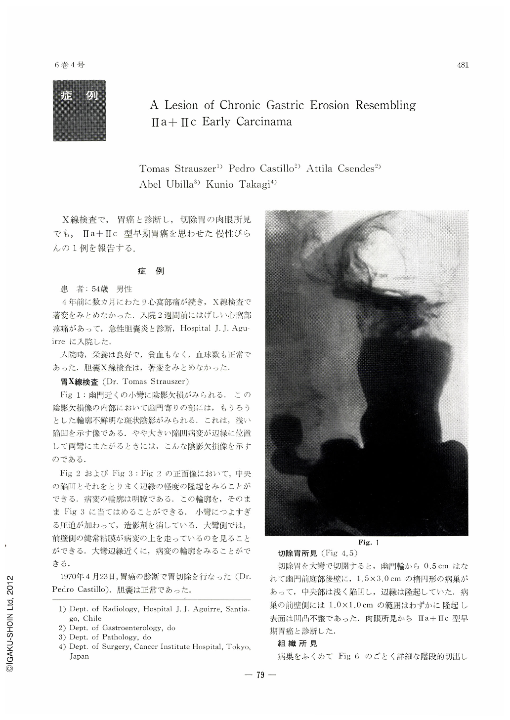
X-Ray Examination of the Stomach (Dr. Tomas Strauszer):
Fig. 1 : A shadow defect is seen on the lesser curvature near the pylorus. Blurred, ill-defined barium flecks, bespeaking of shallow depressions, are observed more distally inside the shadow defect.
Fig. 2 and 3: In figure 2, a frontal view, a central depressed area as well as surrounding, slight marginal elevation is to be seen. The lesion is well-defined. In figure 3, too much pressure has pressed barium away from the lesser curvature. In the greater curvature side, normal mucoral patterns of the anterior wall are superimposed on the lesion. Its contour can be seen near the edge of the greater curvature.
A partial gastrectomy was performed on April 23, 1970, with the diagnosis of gastric carcinoma, whereupon the gall-bladder was found normal in all respects (Dr. Pedro Castillo).
Findings of the sesected specimen of the stomach disclosed an elliptical lesion, 1.5×3.0 cm, on the posterior wall of the prepyloric region (Fig. 4, 5). The edge was elevated and the center shallowly depressed. The area extending within a sphere of 1.0×1.0cm on the anterior wall side of the lesion was found slightly elevated and the surface was uneven and irregular. The case was diagnosed as Ⅱa+Ⅱc early gastric carcinoma on the basis of these gross findings.
Microscopic Findings: Detailed tissue sections were cut from the resectecl gastric specimen, including the lesion, as can be seen from the figure 6 (Dr. Takagi). As shown in Fig. 7, 8, the grossly depressed area was found to be a healed superficial erosion covered with the indifferent epithelium proliferating from the neck part of gland although the depressed area was still erosive in part. Hyperplasias of the foveolar epithelium and of the lamina muscularis mucosae were evident with the grossly elevated mucosa surrounding the depression crater. However, there was no evidence of malignancy.
SUMMARY & CONCLUSION
The case described herein represents one of the types of erosive lesions of the stomach which have been studied and reported by numerous investigators since the first description by Puhl. Usual multiple lesions, though large in size, render diagnostic differentiation from early gastric carcinoma practicable, whereas it is difiicult to distinguish it from Ⅱa+Ⅱc early carcinoma of the stomach when only a single lesion is characterized by depression with a prominent marginal elevation. The subject case had thus been diagnosed as gastric carcinoma from the X-ray and gress finding. One of the authors (Dr. Takagi), too, experienced a case of single gastric erosion, 1,5×2.3cm, as shown in Fig. 9, of which the X-ray and gross findings suggested a Ⅱa+Ⅱc early carcinoma but the subsequent microscopic examination revealed the lesion to be simply an erosion as in the case herein described. Gastric biopsy or cytology, therefore, is eventually necessary in such cases as these, depending upon the lesion encounterecl.
The authors wish to thank Professor Hikoo Shira kabe of Juntendo University for his invaluable in struction of X-ray findings.
Copyright © 1971, Igaku-Shoin Ltd. All rights reserved.